Locations:

Research supported by a recent $3.16 million NCI grant
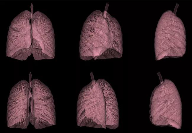
Image content: This image is available to view online.
View image online (https://assets.clevelandclinic.org/transform/cdddc841-5f5e-4765-bbad-1495572498e0/16-PUL-3259-Mazzone-Hero-Image-650x450pxl-jpg)
Lung imaging
A computer-extracted feature analysis of digitalized tissue imaging could soon help identify patients with early stage non-small cell lung cancer (NSCLC) who are likely to face early recurrence after surgery.
Advertisement
Cleveland Clinic is a non-profit academic medical center. Advertising on our site helps support our mission. We do not endorse non-Cleveland Clinic products or services. Policy
With this information, oncologists could more accurately determine which lung cancer patients should undergo aggressive post-surgery chemotherapy and identify patients who could benefit from alternate strategies like immunotherapy to improve chance of cure after surgery.
“Current standard practice includes surgery for most early-stage lung cancer patients,” says Vamsidhar Velcheti, MD, Associate Director for Immuno-Oncology Research, Cleveland Clinic Cancer Center. “Post-surgery chemotherapy can provide added benefit for some patients, but right now we don’t have tests to identify who could benefit.”
The National Cancer Institute recently awarded Dr. Velcheti and Anant Madabhushi, PhD, a $3.16 million grant to advance this promising research project.
“What we’ve proposed with this grant is to create the first actual predictive analysis for early-stage lung cancer to figure out who is going to benefit from chemotherapy,” says Dr. Madabhushi, founding Director of the Center for Computational Imaging and Personalized Diagnostics at Case Western Reserve University School of Engineering. “We haven’t yet definitively shown that it’s predictive, because we haven’t done clinical trials. That is what this new grant will help us to do.”
Dr. Velcheti, Dr. Madabhushi and six collaborators published research regarding the technique in late 2017 in Nature’s Scientific Reports. This retrospective study showed that using a computational histomorphometric image classifier of digitized tissue Hematoxylin and Eosin (H&E) microarray slides could predict recurrence.
Advertisement
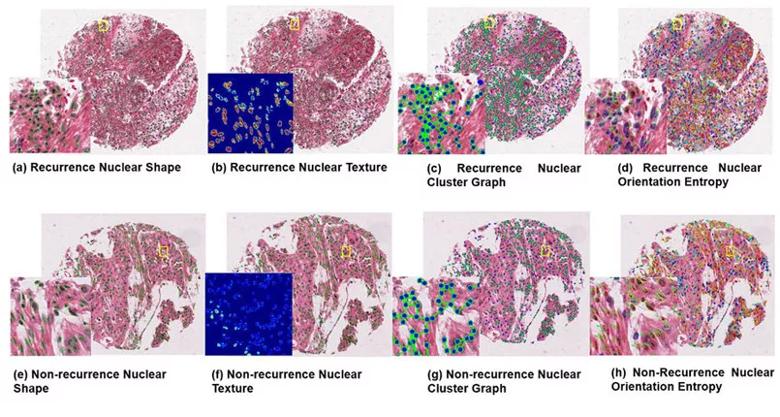
Image content: This image is available to view online.
View image online (https://assets.clevelandclinic.org/transform/3de71ad7-b6b6-45e5-bc60-9fbf20d771a0/velcheti-inset_805x430_jpg)
Caption: Representative TMA tissue spots of recurrent (top row) and non-recurrent (bottom row) NSCLC with corresponding feature maps: Recurrence TMA with (a,e) nuclear shape feature, (b,f) texture feature map (Haralick standard deviation intensity correlation), (c,g) nuclear cluster graph feature map, and (d,h) nuclear orientation. Image republished from: Wang X, Janowcyzk A, Zhou Y, Thawani R, Fu P, Schalper K, Velcheti V, Madabhushi A. Prediction of recurrence in early stage non-small cell lung cancer using computer extracted nuclear features from digital H&E images. Scientific Reports. 2017;7:13543. doi:10.1038/s41598-017-13773-7
The classifier uses computer-assisted digital analysis to identify known clinicopathologic clues, such as tumor size and nuclear morphometric features, of recurrence in early NSCLC to quantify risk. The next step is to show explicitly that the approach can also identify patients who will receive added benefit from adjuvant chemotherapy.
“If this test can identify patients who have a high risk of cancer recurrence, that means other patients may potentially be spared the side effects of chemotherapy,” Dr. Velcheti says.
Advertisement
Advertisement

Creating a safe space for patients

Long-term immune effects reshape preventative strategies and timelines

Large-scale database also reveals potential for immunotherapy to protect against cancer

Findings may help guide discussions around prognosis and allogeneic stem cell transplantation

Research underscores the importance of access to timely diagnosis and treatment in this patient population.

A Cleveland Clinic model combining clinical staging, genomics and AI predicts survival with 18% greater accuracy — and could help match patients to more effective treatments.

Study serves as ‘cautionary tale’ for physicians tempted to rely on liquid biopsy results alone

Direct delivery of viral-based vector KB707 to the lungs may boost anti-tumor response and help overcome immune checkpoint inhibitor resistance