Locations:

Model facilitated understanding of organ’s flaws
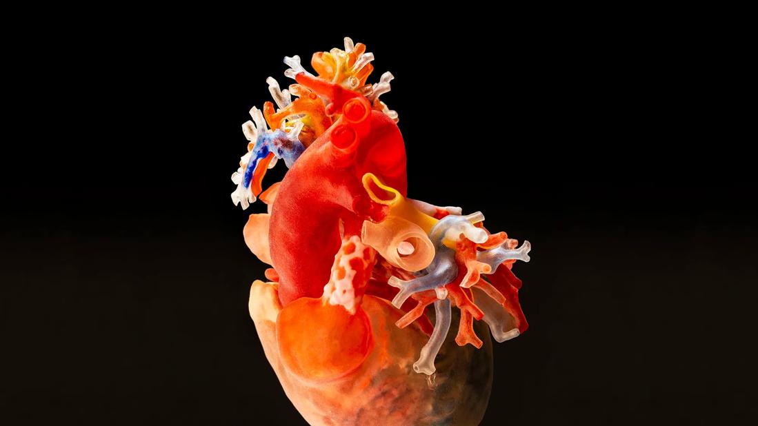
Image content: This image is available to view online.
View image online (https://assets.clevelandclinic.org/transform/1866cb5b-495e-48a1-81a0-fa9a9b25949e/HVI-1572623_04-29-19_018_MLC-jpg)
3D heart
When a 10-year-old Saudi Arabian boy came to Cleveland Clinic last winter with a complicated cardiovascular anatomy due to a congenital defect, the pediatric heart team decided that printing out a three-dimensional (3-D) replica of his heart was the only way they might be able to save it.
Advertisement
Cleveland Clinic is a non-profit academic medical center. Advertising on our site helps support our mission. We do not endorse non-Cleveland Clinic products or services. Policy
The boy’s heart had a hole between the left and right ventricles and some of his valves and ventricles were too small. In addition, his aorta connected to the right ventricle, the chamber that pumps oxygen-poor blood to the lungs, instead of to the left ventricle, the chamber that that pumps oxygen-rich blood to the body.
The child had undergone three past open-heart surgeries. The most recent was a Fontan procedure in which blood from the inferior vena cava and superior vena cava is diverted directly to the pulmonary arteries instead of first flowing through the right ventricle and being pumped by the heart into the pulmonary arteries. This is called a univentricular track because afterward, both ventricles, instead of just the left, pump blood back out to the body.
“Previous surgeons decided the problems inside the heart were too complicated so they decided to leave the inside alone and just alter the outside anatomy,” says Hani Najm, MD, Chief of Pediatric and Congenital Surgery, Cleveland Clinic Children’s.
The Fontan procedure that was performed on the boy worked for a while but eventually it began to fail. The passive blood flow to the lungs meant that blood sometimes backed up into his liver and gastrointestinal tract. Eventually the child began suffering from protein-losing enteropathy and his doctors decided the only way to save him was through a heart transplant.
Because a heart transplant patients often have to wait a long time and because such procedures are not considered a cure, Dr. Najm and his colleague Elizabeth Saarel, MD, Chair of Pediatric Cardiology, Cleveland Clinic Children’s, wondered if they might use 3-D printing technology to better understand the boy’s unique heart anatomy and how to fix it.
Advertisement
“All patients get standard imaging technology, which is an echocardiogram, an MRI and a CT scan and then we have to decide if these are enough to do the repair,” says Dr. Najm. “Ninety-nine percent of the time they would be enough, but in a few cases we cannot see enough from a two-dimensional image—even if it’s 3-D reconstructed on the screen.”
Just as an inkjet printer reproduces a digital image with ink and paper, a 3-D printer reproduces a digital model—in this cased derived from a CT scan—with resin, thermoplastics, photopolymers or other materials. “The printer make an exact replica of the heart, even size-wise,” says Dr. Najm. “Just like when you write a document and send it the printer and it comes back exactly word for word, a 3-D printer takes a 3-D image and lays down these materials instead of ink that are an exact replica.”
Once they had a model of the boy’s heart, Dr. Najm could examine it from every angle and cut it open to see the inside and plan what he might do surgically to repair it.
“With the 3-D model, Dr. Najm was able to see the chambers of the boy’s heart in a 3-D space,” says Dr. Saarel. “He could where they were in relation to one another. He could see the hole inside the boy’s heart. This allowed Dr. Najm to come up with novel connections that were tailored to that particular patient.”
Dr. Najm ended up performing something close to what’s known as a double switch. He switched the aorta to its proper place—the left ventricle. Then using the hole between the ventricles, he connected the inferior vena cava to the pulmonary arteries. This allowed blood from the lower part of the body to pass through the right ventricle and be pumped into the lungs so there would no longer be blood backing up into the liver and gastrointestinal tract. He left the superior vena cava—which receives blood from the head and neck—connected directly to the pulmonary arteries.
Advertisement
“We executed the operation exactly as we had planned it with the 3-D replica,” says Dr. Najm. “The child ended up improving dramatically. He went home10 days after surgery and he was eating well and playing and obviously he went home with his own heart not a heart transplant.”
He adds: “Without 3-D printing, I could not have done this operation.”
Watch this video with Dr. Najm for more about this case.
Advertisement
Advertisement

Cleveland Clinic welcomes world-leading chest wall surgeon Dr. Hyung Joo Park

Common misconceptions about perioperative management and strategies to improve care

Life-changing gene therapy is currently under FDA review

How evolving medical therapies and more precise patient selection are reshaping the role of intestinal transplantation

Cleveland Clinic specialist discusses the new clinical practice guideline

Introducing Prajwal Rajappa, MD, MS, Director of Pediatric Precision Oncology

The one-stop THRIVE clinic addresses renal, psychosocial, educational and other challenges alongside cardiac surveillance

Study found lower rates of nausea, vomiting and healthcare use through 12 months of follow-up