Locations:

Study compares outcomes of minimally invasive interval debulking and laparotomy
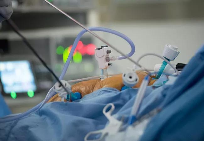
Image content: This image is available to view online.
View image online (https://assets.clevelandclinic.org/transform/14a862e6-ae5d-4765-95e7-f386baf5b3f0/HIPEC_650x450_jpg)
HIPEC_650x450
Hyperthermic intraperitoneal chemotherapy (HIPEC) performed at the time of interval debulking surgery (IDS) offers life-extending benefits to patients with epithelial ovarian cancer that has spread to the abdominal cavity. At Cleveland Clinic, gynecologic oncologists have combined minimally invasive interval debulking surgery (MIIDS) with the life-extending benefits of HIPEC for appropriate candidates.
Advertisement
Cleveland Clinic is a non-profit academic medical center. Advertising on our site helps support our mission. We do not endorse non-Cleveland Clinic products or services. Policy
In patients with epithelial ovarian cancer, HIPEC with single-agent cisplatin at the time of cytoreductive surgery has been shown to increase overall survival by 12 months compared with interval cytoreductive surgery, with no increase in perioperative morbidity. HIPEC is usually done after optimal debulking as part of an exploratory laparotomy.
With the minimally invasive technique performed at Cleveland Clinic, HIPEC can be performed during interval debulking, reducing length of stay with no increase in perioperative morbidity. “We’re one of a small number of centers in the country using HIPEC and perhaps the only center using HIPEC in the minimally invasive setting for these cancers,” says Cleveland Clinic’s Molly Morton, MD.
In a retrospective study led by Chad Michener, MD, Medical Director of Continuous Improvement for Cleveland Clinic’s Women’s Health Institute, peri-operative outcomes were compared in women with advanced ovarian cancer who underwent HIPEC at the time of interval debulking through either minimally invasive or open approaches. Among the 10 patients with ovarian cancer who underwent HIPEC, the median length of stay was shorter in patients who had minimally invasive surgery with HIPEC compared to laparotomy. There were no differences in major and minor complications between the groups. In six cases, HIPEC was done through a single port; of the remaining two cases, one was laparoscopic and the other robotic. In all 10 patients who underwent minimally invasive procedures, optimal debulking to no gross residual disease was achieved.
Advertisement
“Overall, patients who had this type of surgery had similar cytoreduction and tolerated the surgery very well,” according to Laura Chambers, DO. “Previously, HIPEC candidates faced invasive surgery. Our study indicates that HIPEC can be performed in the setting of MIIDS, and that MIIDS achieves similar rates of debulking. Future research will look at survival outcomes.”
“Although using this technique for debulking surgery is relatively new, we have used it since 2016 for initial staging of newly diagnosed gynecologic cancers that have a propensity for intra-abdominal spread,” says Dr. Michener.
The technique begins with manual palpation of the entire peritoneal cavity to assess disease distribution and appropriateness for the minimally invasive approach. Dr. Michener emphasizes the importance of this step, noting that if it is omitted, disease that is not easily seen with laparoscopic visualization alone may be missed.
Once cytoreduction is complete, inflow and outflow tubing is placed via the single port incision. The falciform ligament may be transected using a bipolar vessel sealing device to facilitate correct placement of the HIPEC tubing. The umbilical incision also may be extended 1 to 2 cm to accommodate the tubing.
Says Dr. Michener, “A 7- to 8-cm incision is utilized for the palpation of the entire abdominal cavity. This is followed by a hybrid approach of direct resection of a portion of the omentum through the incision as well as single-port laparoscopic removal of residual disease.”
Advertisement
The looped outflow tubing is introduced into the abdomen and placed in the upper quadrants over the liver bed and along the diaphragm. The bifurcation of the inflow Y tubing is placed over the outflow tubing and into the lower quadrants and pelvis.
To distinguish the tubing, the inflow tubing has red arrows pointing toward the abdomen and the outflow tubing has blue arrows pointing away from the abdomen. The color coding also correlates with temperature.
Once temperature probes are connected to the HIPEC machine, the fascia are closed and the inflow and outflow tubing secured starting at the cranial apex with sutures. A water-tight seal of the fascia and skin, and any accessory ports, is necessary to prevent spillage of HIPEC during infusion.
At Cleveland Clinic, the Thermocam HT2000 device is used to deliver HIPEC (cisplatin 100 mg/m2 with or without Taxol135-175mg/m2) to the abdominal cavity as per standard protocol. The solution is circulated for 90 minutes with a goal outflow temperature of 42°C.
After HIPEC is complete, the patient’s abdomen is irrigated with normal saline via tubing and the fluid is evaluated. The tubing is removed and the incision closed.
“The hand-assist minimally invasive debulking allows for tactile assessment of disease within the entire peritoneal cavity while also allowing us to complete HIPEC through a relatively small incision,” concludes Dr. Michener. “It also allows for flexibility of the incision size, which can be useful for extraction of larger masses as well as for intestinal surgery.” Trials are underway comparing progression free survival for women undergoing MIIDS with HIPEC compared to an open debulking procedure.
Advertisement
Advertisement

Research findings offer clues for improving disease outcomes in men

Creating a safe space for patients

Long-term immune effects reshape preventative strategies and timelines

Large-scale database also reveals potential for immunotherapy to protect against cancer

Findings may help guide discussions around prognosis and allogeneic stem cell transplantation

Research underscores the importance of access to timely diagnosis and treatment in this patient population.

A Cleveland Clinic model combining clinical staging, genomics and AI predicts survival with 18% greater accuracy — and could help match patients to more effective treatments.

Study serves as ‘cautionary tale’ for physicians tempted to rely on liquid biopsy results alone