Locations:

Cleveland Clinic specialists discuss challenges and opportunities for managing the disease
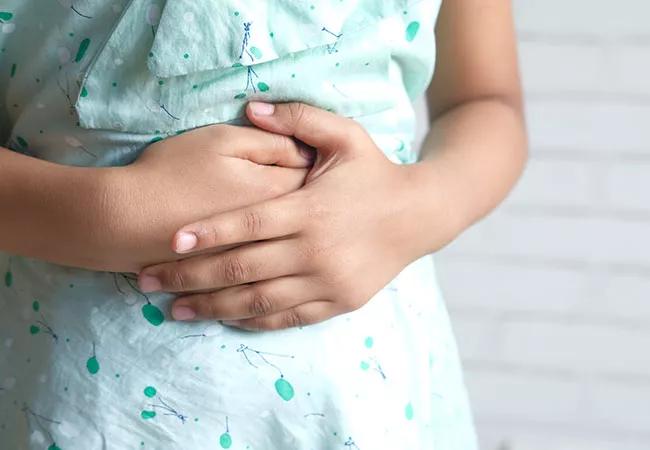
Image content: This image is available to view online.
View image online (https://assets.clevelandclinic.org/transform/2cb27927-3e83-4157-a51b-355ef5e7433e/23-CHP-3560021-CQD-Kumar-Daniels-CME-Live-3_jpg)
23-CHP-3560021-CQD–Kumar-Daniels—CME-Live-3
Nonalcoholic fatty liver disease (NAFLD) is the leading cause of liver disease in children, and NAFLD-related cirrhosis is the leading cause of liver transplants in young adults. Nearly 10% of children between ages 2 and 19 have NAFLD, with up to 38% of children with obesity in the United States affected.1
Advertisement
Cleveland Clinic is a non-profit academic medical center. Advertising on our site helps support our mission. We do not endorse non-Cleveland Clinic products or services. Policy
According to three Cleveland Clinic Children’s experts who spoke at a recent continuing medical education event, the challenges for clinicians include delayed diagnoses because of inconsistencies in testing and interpretation, effective management of childhood obesity and inadequacy of data applicable directly to pediatric patients.
On the positive side, liver fibrosis has been shown to be a sensitive histologic marker, recommendations are being developed for interpretation of alanine aminotransferase (ALT) levels in pediatric patients, and epidemiologic data specific to pediatric patients are accumulating.
“NAFLD is a burden on American children’s future health,” says Vera Hupertz,MD. “This is a health crisis with multiple contributors that requires multidisciplinary management.” Dr. Hupertz is the Medical Director of the Pediatric Liver Care Center at Cleveland Clinic Children’s.
There are three sets of guidelines for evaluation of possible NAFLD in children with obesity and overweight:
The American Academy of Pediatrics (AAP) recommends checking fasting lipids, fasting glucose and ALT every two years starting at age 10 among children with obesity, defined as body mass index (BMI) above the 95th percentile. For children who are overweight (BMI at or above the 85th percentile and below the 95th percentile) the AAP recommends only a fasting lipid panel if the child is free of risk factors. The AAP does not recommend tests for abnormal glucose metabolism or liver function for children who are overweight and under age 10. The North American Society for Pediatric Gastroenterology, Hepatology, and Nutrition (NASPGHAN) guidelines parallel those of the AAP; i.e., monitor ALT in the presence of obesity.
Advertisement
The European Society for Pediatric Gastroenterology, Hepatology, and Nutrition (ESPGHAN) guidelines recommend that all children with obesity or overweight be screened starting at age 3 with ALT and ultrasound.
In a retrospective chart review, Ezaizi et al2 evaluated the guidelines in 344 patients, median age 13, referred to a pediatric hospital-based weight management program. The current NASPGHAN guidelines identified 20% of patients with NAFLD. When ultrasound was considered, as required by the ESPAGHAN guidelines, 58% were identified.
Dr. Hupertz says that a screening strategy combining elevated ALT and fatty infiltration observed on ultrasound will enhance the sensitivity of detection of NAFLD in at-risk children.
ALT is used in pediatric screening, but the threshold for detecting liver disease is uncertain. In addition to variations in “normal,” laboratories do not exclude patients who may have underlying liver disease such as NAFLD from population analyses. This skews the distribution levels of ALT upward, setting the upper limit of normal falsely high.
“If this is not recognized, especially in children,” cautions Dr. Hupertz, “we risk missing potentially treatable liver disease or exposing young children to unnecessary risks with hepatotoxic medications.”
Dr. Hupertz warns that children with overweight who have central obesity, signs of insulin resistance, prediabetes or diabetes, dyslipidemia, or sleep apnea are at high risk for developing NAFLD, and adolescents with severe obesity are more likely to present with advanced liver disease and severe systemic inflammation.
Advertisement
“Concerns about overtesting and cost are legitimate,” she says, “but the impact of obesity and comorbidities on the long-term health and mortality of young patients is an equal concern.”
Liver biopsy is the gold standard for assessment of suspected NAFLD, given its ability to evaluate different aspects of the disease including steatosis, nonalcoholic steatohepatitis (NASH) activity and fibrosis stage, says Mohammad Nasser Kabbany, MD. But, he notes, there is increasing use and study of noninvasive imaging methods and growing potential for biomarkers. Dr. Kabbany practices pediatric gastroenterology, hepatology, and nutrition at Cleveland Clinic Children’s.
Noninvasive imaging methods to evaluate steatosis
Advertisement
Biomarkers for NASH can potentially enhance diagnosis, but they have not been studied extensively in children.
Cytokeratin 18 (CK18), a result of apoptosis of hepatocytes by caspase 3, is one of the most widely investigated biomarkers, but a test to measure it is not commercially available.
Liver enzyme measurement, an inexpensive point-of-care test, sometimes correlate clinically with histologic improvement. In a clinical trial of cysteamine therapy in pediatric patients with biopsy-confirmed NAFLD, histologic improvement correlated with improved ALT, aspartate transaminase (AST), and gamma-glutamyl transferase.4 The strategy is limited by the lack of an ALT or AST cutoff that indicates the presence or absence of NASH.
“The bottom line is that none of the currently available serum markers and imaging modalities have the ability to differentiate NASH from simple steatosis with high sensitivity and specificity,” says Dr. Kabbany.
Several histologic scoring systems have been used to stage fibrosis in chronic liver diseases including NAFLD. Most of them range from F0 (no fibrosis) to F4 (cirrhosis). Advanced fibrosis is defined as F3 (bridging fibrosis). Studies involving adults have shown significant correlation between advanced fibrosis and morbidity and mortality.
Liver stiffness measurement (LSM) can be generated by VCTE. The LSM varies depending on body habitus, the type of probe used, and the fasting state.
Normal LSM for pediatric patients ranges from 3.4 to 5.1 kPa. Several pediatric studies showed that LSM correlates well with the stage of fibrosis. However, most pediatrics studies have included not only patients with fatty liver, but also patients with other chronic liver diseases which makes it challenging to come up with accurate cut-off values as different etiologies may have different LSM cutoffs. Further, adds Dr. Kabbany, most studies in children are based in tertiary referral centers, limiting their application to the general population. LSM measured via VCTE is a promising noninvasive tool, but further studies are needed to better define cut-offs to stage fibrosis. Pediatric guidelines have not endorsed VCTE for ruling out advanced fibrosis.
Advertisement
Acoustic radiation force impulse, an ultrasound-based fibrosis staging tool, provides results that correlate with liver stiffness measured on biopsy. It has the advantage to be more successful in morbidly obese patients. Studies in pediatric patients are scarce.
Magnetic resonance elastography generates an elastogram, a global assessment of liver stiffness usually measured in kPa. As with other MRI-based studies, insurance coverage is a challenge, as is use in small children.
“The bottom-line is noninvasive tools are gaining momentum in assessing different aspects of NAFLD. More studies are needed in children to validate them,” concludes Dr. Kabbany. “Until then, liver biopsy remains the gold standard to assess various aspects of this growing pandemic.”
“Liver fibrosis is the most sensitive histologic marker associated with poor outcomes,” says Mike Leonis, MD, PhD. Dr. Leonis is a Staff Physician in the Department of Pediatric Gastroenterology, Hepatology, and Nutrition and Medical Director of Pediatric Liver Transplantation at Cleveland Clinic Children’s.
Insights from NASH Clinical Research Network
Findings from the NASH Clinical Research Network (CRN) support the significance of fibrosis. The NASH CRN, comprising eight adult and nine pediatric sites in the United States, performed a prospective study of outcomes in 1,700 patients with NAFLD with a mean follow-up of four years.6 Analyses have shown that F3 and F4 fibrosis correlate with hepatic decompensation. As a result, complications of portal hypertension, such as esophageal variceal bleeding, are greater with F3 or F4 fibrosis, and risk of hepatocellular carcinoma is markedly increased.
What have we learned about pediatric NAFLD?
Although children are less represented in the NASH CRN, some pediatric data have been gathered:
A recent study by Draijer et al9 examined the natural history of pediatric NAFLD over a 10-year-period in 133 children with severe obesity. Of the original cohort, 38% were available for follow-up 10 years later. The investigators used MR spectroscopy to assess steatosis and the enhanced liver fibrosis test to assess fibrosis.
At baseline, on biopsy, 24 patients had NAFLD and 27 had no NAFLD; 35% had dyslipidemia and none had type 2 diabetes. At follow-up 10 years later, 30% of those who had NAFLD at baseline had resolution of steatosis, and 30% of those who had no NAFLD at baseline had developed NAFLD.
“The jury is still out on what to do,” says Dr. Leonis. “Although we see both improvement and worsening, who will be in which group remains unpredictable. Many children develop advanced fibrosis, and we need to figure out how to rescue these children, so they don’t end up needing liver transplants in the future.”
References
Advertisement

Research demonstrates cirrhosis regression in one-third of patients, with higher rates using alternative assessment

Multidisciplinary framework ensures safe weight loss, prevents sarcopenia and enhances adherence

Findings could help clinicians make more informed decisions about medication recommendations

Findings are first to provide underlying explanation, linking the diagnosis to high-immune activation and worse clinical outcomes
Histotripsy is noninvasive and may generate abscopal effect

IBAT inhibitors, elastography and more
New device offers greater tumor control for malignant liver lesions
Reviewing how the drug can be incorporated into care