Locations:

Brief pearls for diagnosis and management of ascites and relevant conditions associated with decompensated cirrhosis

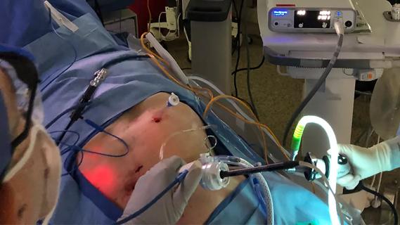
Image content: This image is available to view online.
View image online (https://assets.clevelandclinic.org/transform/9f02cd7d-c83d-49b1-b58d-be75a4ee7a74/23-DDI-4107008-CQD-650x450-1_jpg)
Liver disease
Written by Sarah Kahn, MD, and Maureen Linganna, MD
Advertisement
Cleveland Clinic is a non-profit academic medical center. Advertising on our site helps support our mission. We do not endorse non-Cleveland Clinic products or services. Policy
Note: This article is reprinted from the Cleveland Clinic Journal of Medicine (2023;90[4]:209-213).
In response to significant advances of antimicrobial resistance and studies comparing therapeutic options for ascites and hepatorenal syndrome, the American Association for the Study of Liver Disease published a new 2021 guidance1 as a comprehensive guide for both outpatient and inpatient diagnostic evaluation and management of ascites, updated information regarding use of albumin, and specified definitions and management recommendations for hyponatremia.
Development of ascites is associated with a reduction of 5-year survival from 80% to 30%,1,2 largely associated with complications that include infection and hepatorenal syndrome. A thorough evaluation is required for diagnosis of new ascites to exclude other etiologies, including heart failure, renal failure, infections or malignancy.1 Complete initial analysis should consist of laboratory evaluation, abdominal Doppler ultrasonography and a diagnostic paracentesis,1 although no data currently support this recommendation. A serum ascites albumin gradient 1.1 g/dL or greater suggests portal hypertension, massive liver metastases or right heart failure.1,3 In addition to patients with symptoms suggestive of infection (e.g., fevers, abdominal pain), ascitic fluid cultures should be obtained for any decompensating patient, including for the development of encephalopathy, acute kidney injury or jaundice.1
In general, angiotensin II receptor-antagonists, angiotensin-converting enzyme inhibitors and non-steroidal anti-inflammatory drugs should be avoided in patients with ascites owing to impact on effective circulating volume and renal perfusion.1 Though not directly nephrotoxic, use of angiotensin-converting enzyme inhibitors or angiotensin II receptor-antagonists was noted to correlate with increased risk of end-stage renal disease in cirrhotic patients with ascites.4
Advertisement
Based on treatment response, ascites can be classified as responsive, recurrent or refractory.5 Initial management of ascites includes 2 g sodium restriction. Diuresis initially with spironolactone 100 to 200 mg daily is suggested for first-time ascites, and dose adjustments should be made at intervals of at least 72 hours, up to a maximum daily dose of 400 mg.1 For recurrent ascites, combination therapy with furosemide and spironolactone is recommended with a starting dose of 40-mg furosemide to a maximum 160-mg dose daily.5 Once ascites has been mobilized, diuretics should be tapered to the lowest effective dose to minimize adverse effects.1 In some cases (approximately 5% to 10% of all patients with cirrhosis), ascites cannot be managed medically and becomes refractory, with 50% survival at six months.1,6
Refractory ascites occurs when one of three criteria are met: recurrence as grade 2 or 3 within four weeks of mobilization with diuretic therapy (early recurrence),1,7 persistence despite maximum diuretic dosage (diuretic resistant) or recurrence or persistence of side effects from attempting to increase diuretics (diuretic intolerant).1
Therapeutic large volume paracentesis (> 5 L) can be used for refractory ascites with fewer side effects than diuresis.1,8,9 Removal of large amounts of fluid, particularly > 8 L, can lead to circulatory shifts and postparacentesis circulatory dysfunction, which manifests as hepatorenal syndrome, hepatic encephalopathy, or dilutional hyponatremia.1,10,11 Albumin infusion with 6 to 8 g/L of ascitic fluid removed is recommended to mitigate this risk.1,10,12
Advertisement
Nonselective beta-blockers, used in managing portal hypertension, are associated with higher incidence of postparacentesis circulatory dysfunction,13,14 although there is insufficient evidence to recommend against their use in cirrhosis. Instead, caution is advised in the setting of renal insufficiency, hyponatremia or hypotension.15
Transjugular intrahepatic portosystemic shunt placement is a useful treatment for refractory ascites in certain patients, particularly for those with low Model for End-stage Liver Disease scores16 and confers a 93% chance of one-year transplant-free survival compared with 53% for patients managed with paracenteses, diuretics, and albumin.17 Following placement, it may take up to six months for ascites resolution, and so salt restriction should be continued following transjugular intrahepatic portosystemic shunt placement. It is recommended to discontinue diuretic therapy to allow return of splanchnic volume to systemic circulation. Despite good results in patients with low Model for End-stage Liver Disease scores, scores ≥ 18 are generally considered high risk for transjugular intrahepatic portosystemic shunt.18 Patients who are not candidates for transjugular intrahepatic portosystemic shunt should be considered for referral for liver transplantation.1,18
Hyponatremia and hepatic hydrothorax are also frequently encountered with cirrhosis, defined as a serum sodium less than 135 mEq/L.1,19 Seen in 49% of patients with cirrhosis, low serum sodium is associated with severe ascites and frequent ascitic complications.19 The most common subtype is hypervolemic hyponatremia, owing to third spacing and vasopressin activation, while hypovolemic hyponatremia may occur with diuretic use.19,20 Rate of sodium correction is based on acuity with goal rate of increase in serum sodium for chronic cases of 4 to 6 mEq/L over 24 hours.21,22 In acute cases, correction should be faster, though the exact rate is not specified in the guidance.1 Specific management of hyponatremia is based on severity, as follows1,3,17,23:
Advertisement
Hepatic hydrothorax, a difficult-to-manage complication of cirrhosis, is a transudative pleural effusion due to translocation of peritoneal fluid through diaphragmatic defects and reported to occur in 4% to 12% of patients with cirrhosis.24 Though typically right-sided, it may occur on the left, bilaterally, and in the absence of ascites.24 It is associated with high mortality risk, exceeding that predicted by the Model for End-stage Liver Disease score.1,3,24 Management is similar to that of ascites, with fluid restriction and diuresis.1 Abdominal hernias, particularly umbilical hernias, are common in the setting of ascites due to increased intra-abdominal pressure. Surgical repair may be considered when ascites management and nutritional status have been optimized.24
The most common source of bacterial infection in patients with cirrhosis is spontaneous bacterial peritonitis (SBP), accounting for 27% to 36% of infections.1,4,25,26 Clinical deterioration (i.e., jaundice, altered mentation, or acute kidney injury) should prompt exclusion of SBP with a diagnostic paracentesis. In hospitalized patients, diagnostic paracentesis should be performed even in the absence of symptoms suggestive of SBP.27 Diagnosis of SBP is established when the fluid absolute neutrophil count is greater than 250 cells/ mm3 and is further confirmed with positive cultures.1,28 Empiric intravenous antibiotics after cultures are obtained are the mainstay of management of SBP and spontaneous bacterial empyema, as each hour’s delay in treatment increases mortality by 10%.1,4,5,29
Advertisement
Effective empiric antibiotic choice plays a key role in timely management of SBP.1 Third generation cephalosporins are effective if local prevalence of multidrug resistant organisms is low, while broad coverage therapy (i.e., piperacillin-tazobactam with vancomycin) is recommended for high prevalence of multidrug resistant organisms, history of prior multidrug resistant organisms infection, nosocomial and hospital-acquired infections, or in critical illness. Daptomycin should be added if there is history of vancomycin-resistant Enterococcus. Some confusion arises with positive cultures and fewer than 250 cells/mm3 of neutrophils; such cases do not require antibiotics and likely are contaminants.1
In addition to antibiotics, albumin dosed at 1.5 g/kg on day 1 and 1 g/kg on day 3 should be administered and is especially helpful if concomitant acute kidney injury or jaundice are present.5,30,31 Repeat paracentesis/thoracentesis after two days of therapy may be done to assess treatment response.1 Treatment, secondary prophylaxis with norfloxacin, or ciprofloxacin in the absence of norfloxacin, should be used. For cases of gastrointestinal hemorrhage, prophylaxis with intravenous ceftriaxone 1 g every 24 hours for seven days should be used.1 Primary SBP prophylaxis should also be considered in the following cases of cirrhosis without bleeding1:
Patients with cirrhosis and ascites are at risk of acute kidney injury (i.e., increase in creatinine ≥ 0.3 mg/dL within 48 hours or ≥ 50% increase in creatinine over seven days), with an estimated prevalence in hospitalized patients between 27% and 53%.1,32,33 The two most common causes of acute kidney injury are prerenal azotemia and acute tubular necrosis. Prerenal azotemia may be secondary to hypovolemia or hepatorenal syndrome. The diagnosis of hepatorenal syndrome is made once hypovolemia/shock, nephrotoxic exposure, and structural kidney damage have been excluded in a patient with ascites who presents with prerenal acute kidney injury.1,32,33
The principle management of hepatorenal syndrome is vasoconstrictor and albumin therapy for up to 14 days. In the United States, midodrine and octreotide in combination are used for hepatorenal syndrome therapy, though their efficacy is low.33 The preferred treatment is terlipressin, a vasoconstrictor that may be used outside of the intensive care unit, which has been shown to improve the likelihood of reversal of hepatorenal syndrome without dialysis and 10-day survival relative to placebo (29.1% vs 15.8%; P = .012).3,34,35 It was very recently approved in the United States in limited settings, and centers are in the process of developing protocols to incorporate its use for hepatorenal syndrome treatment.36 An alternative with comparable efficacy is norepinephrine, though use is limited to the intensive care unit.1 Some studies have looked into using vasopressin in place of octreotide, which has been associated with improved survival and recovery, though use in the United States has thus far been limited.1,37
Response to therapy may be defined as creatinine decrease to less than 1.5 mEq/L or within 0.3 mEq/L of baseline.1 If a response is not seen on maximum doses of therapy for four consecutive days, vasoconstrictors may be discontinued.1 In treatment failure, renal replacement therapy is reserved for those referred for transplant or based on reversibility of other organ dysfunction. In patients with limited expectations for renal recovery, dual liver-kidney transplant may be considered.
References
Advertisement

Research demonstrates cirrhosis regression in one-third of patients, with higher rates using alternative assessment

Multidisciplinary framework ensures safe weight loss, prevents sarcopenia and enhances adherence

Findings could help clinicians make more informed decisions about medication recommendations

Findings are first to provide underlying explanation, linking the diagnosis to high-immune activation and worse clinical outcomes

Histotripsy is noninvasive and may generate abscopal effect

IBAT inhibitors, elastography and more
New device offers greater tumor control for malignant liver lesions

Reviewing how the drug can be incorporated into care