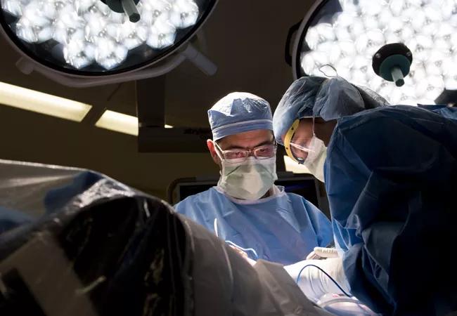
A robust, multidisciplinary team key to best possible outcomes

Restorative proctocolectomy has become the surgical procedure of choice for patients with ulcerative colitis, familial polyposis and selected patients with Crohn’s disease, when indicated. In these cases, a pouch is created when loops of small bowel are folded on themselves, and connected with each other to create a reservoir that can be connected to the anal canal to be used in lieu of the rectum. This maintains the natural route of defecation, allowing patients to pass stools through the anus voluntarily and avoid a permanent stoma.
Advertisement
Cleveland Clinic is a non-profit academic medical center. Advertising on our site helps support our mission. We do not endorse non-Cleveland Clinic products or services. Policy
Sometimes, however, patients develop a complication or have poor function following a pouch operation. These complications include anastomotic leaks, pre-sacral sinuses and pouch-vaginal fistula. Other problems that may significantly impact the pouch function include afferent limb syndrome, pouch twist and refractory pouchitis, in addition to strictures and long rectal cuff/cuffitis.
A pouch salvage procedure, also known as a “redo pouch,” is a “very technically demanding procedure,” says Cleveland Clinic colorectal surgeon Sherief Shawki, MD. “It requires not only a capable surgeon who is familiar with different pouch problems and how to fix them, but a whole team of ancillary service professionals who are familiar with caring for the patient during surgery and afterward on the nursing floor.”
This multidisciplinary approach can involve experts in colon and rectal surgery, gastroenterology, radiology, pathology, pain management and psychology. “We’re treating the patient as a whole and not just ‘the pouch,’” he notes. Once the clinical team identifies the specific etiology, they design a management plan to promote the best possible outcome for each individual.
Surgeons specializing in redo pouches at Cleveland Clinic’s main campus include Sherief Shawki, MD, Tracy Hull, MD, Scott Steele, MD, Stefan Holubar, MD, and Conor Delaney, MD, PhD. At Cleveland Clinic Florida, the team includes Steven Wexner, MD, Dave Maron, MD, and Giovanna da Silva Southwick, MD.
Nonsurgical approaches are used when possible through collaboration with gastroenterologist Bo Shen, MD, who is internationally recognized for endoscopic management of pouch complications and established the Pouchitis Clinic in 2004. Drs. Shen and Hull recently established a first-of-its-kind IBD board to help reach consensus on the most complex cases.
Advertisement
As an example, 48-year-old woman who underwent ileal pouch anal anastomosis and diverting loop ileostomy elsewhere, presented with severe pain and liquid stool coming out her vagina. Following a comprehensive history and review of the external hospital operative records, the patient underwent examination under anesthesia. Dr. Shawki discovered bile in the vagina, pouch-anal-vaginal fistula (connection) and pre-sacral track, without overt sepsis. Dr. Shawki and team initially sutured closed the limb of the loop ileostomy going towards to the pouch to control the pouch vaginal fistula.
Eight months later, the patient returned to the operative room, and the old pouch was excised from the pelvis. Because her anal sphincters were intact and functional, Dr. Shawki created a new pouch anal anastomosis after rotating the mesentery of the pouch anteriorly to minimize the risk of fistulization with the repaired vagina. At the same time, they also created a new diverting loop ileostomy.
Recently the loop ileostomy was reversed, the patient was able to use her pouch again, and she avoided having to wear a bag forever.
Another patient, a 35-year-old woman with a “J pouch,” presented with difficulty defecating since her ileostomy reversal. Dr. Shawki and team took a thorough history, performed an endoscopic exam and suspected afferent limb syndrome. In the operating room, they discovered the small bowel loops just proximal to the pouch folded and kinked in the pelvis anterior to the pouch. They meticulously divided adhesions, released the loops and performed surgery to minimize recurrence of the problem without the need to create ileostomy. Afterwards, the patient reported significant improvement in her bowel function.
Advertisement
“Overall, any problem that impairs the inflow, outflow or integrity of the pouch body can result in pouch malfunction,” Dr. Shawki says. “Effectively resolving these issues, again, requires a team familiar with these issues as well as with this patient population.”
Patient management generally includes a thorough history and physical examination and evaluation of the pouch body orientation, inflow, outflow and integrity of the suture lines, and obtaining biopsies under anesthesia. Endoscopy is usually performed as well.
Imaging also plays an important role in diagnosis. For example, a contrast enema can outline the pouch configuration and help determine whether there are any leaks. Clinicians often use MRI to look for occult sepsis, fistula and/or sinuses. Ultimately, tissues are sampled for histopathology evaluation.
“Collectively, we come together as a multidisciplinary team that includes gastroenterologists, radiologists and pathologists, with the colorectal surgeon as the lead to make the best decisions to help ensure the best outcomes,” Dr. Shawki says.
Advertisement
Advertisement

Multidisciplinary framework ensures safe weight loss, prevents sarcopenia and enhances adherence

Study reveals key differences between antibiotics, but treatment decisions should still consider patient factors

Key points highlight the critical role of surveillance, as well as opportunities for further advancement in genetic counseling

Potentially cost-effective addition to standard GERD management in post-transplant patients

Findings could help clinicians make more informed decisions about medication recommendations

Insights from Dr. de Buck on his background, colorectal surgery and the future of IBD care

Retrospective analysis looks at data from more than 5000 patients across 40 years

Surgical intervention linked to increased lifespan and reduced complications