Locations:

A case report of a thumbnail deformity
Editor’s note: This is an abridged version of an article originally published in the Cleveland Clinic Journal of Medicine. The article in its entirety, including a complete list of references, can be found here.
Advertisement
Cleveland Clinic is a non-profit academic medical center. Advertising on our site helps support our mission. We do not endorse non-Cleveland Clinic products or services. Policy
By Taylor Bullock, BS, Daniel Michalik, MD, Rashmi Unwala, MD, and John Anthony, MD
A 65-year-old man presented with a thumbnail deformity that had been present for three months. He said he had no pain and no history of trauma to the nail.
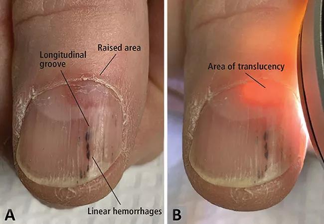
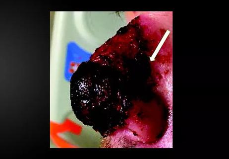
Examination of the thumbnail revealed a longitudinal groove, distal linear hemorrhages and a raised area at the proximal end that appeared to be a subungual nodule affecting the underside of the nail plate, and transillumination showed a translucent area 3 mm by 5 mm beneath the nail matrix. Examination of all other digits was unremarkable.
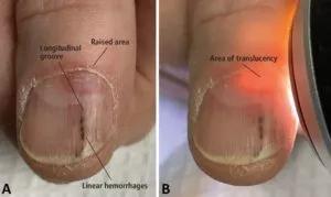
Image content: This image is available to view online.
View image online (https://assets.clevelandclinic.org/transform/694daf1c-257a-42fd-8c91-1f85fcf9f0ce/subungual-300x179_jpg)
Subungual digital mucous cyst of the thumbnail: (A) longitudinal groove, distal linear hemorrhages and a raised area at the proximal nail; and (B) a translucent area, 3 mm × 5 mm, beneath the nail matrix.
Clinically, this presentation appeared to be consistent with a subungual digital mucous cyst (DMC) causing nail deformity due to compression of the nail matrix. To confirm the diagnosis, a sterile 22-gauge needle was used to drill through the nail plate into the translucent area. A copious amount of clear, gelatinous material, characteristic of a DMC, was expressed from the puncture site.
Longitudinal grooves in the nail bed can be caused by median nail dystrophy, trauma, compression of the nail matrix from tumors and physiologic furrows and ridges that are accentuated in diseases such as lichen planus and Darier disease. Tumors that can affect the nail matrix include various nail fibroma, DMC, pyogenic granuloma, glomus tumor, subungual exostosis, squamous cell carcinoma and melanoma.
Advertisement
The translucency of this patient’s subungual nodule was highly suggestive of a DMC, and the clear, gelatinous material expressed from the cyst confirmed it. In addition, a red lunula, as seen in our patient, is a common finding in subungual DMC.
DMC, also known as myxoid pseudocyst or synovial cyst, commonly presents as a superficial, dome-shaped, shiny, cystic nodule located near the distal interphalangeal joint on the dorsum of the fingers. The cyst is commonly diagnosed clinically based on the appearance and the history of intermittent discharge of a mucoid substance. DMC is more common in people with osteoarthritis and in women.
While superficial DMC is common, the prevalence of subungual DMC is unknown, as few have been reported. The focal collection of fluid in DMC lacks a cystic lining, so DMC is not a true cyst. DMC can cause nail deformities by compressing nail matrix cells, most commonly proximal and superficial to the proximal nail fold.
Subungual DMC is much more difficult to diagnose than its superficial counterpart, not only because it is less accessible, but also because it lacks the characteristic appearance of superficial DMC and does not cause intermittent mucinous discharge. Though DMC is commonly asymptomatic when left untreated, subungual DMC most often requires nail avulsion and surgery to rule out other subungual masses, including malignancies.
Transillumination is a quick and easy way to diagnose DMC and rule out other subungual masses, as seen in this case. The finding of a translucent cyst can spare the patient from undergoing nail avulsion and surgery. When evaluating subungual masses, all clinicians should be aware of the utility of transillumination in the diagnosis of subungual DMC.
Advertisement
Mr. Bullock is a medical student at Cleveland Clinic Lerner College of Medicine. Dr. Michalik is a resident in the Department of Dermatology. Drs. Unwala and Anthony are staff in the department.
Advertisement
Advertisement
Family history may eclipse sun exposure in some cases
Consider secondary syphilis in the differential of annular lesions
Persistent rectal pain leads to diffuse pustules

Two cases — both tremendously different in their level of complexity — illustrate the core principles of nasal reconstruction
Stress and immunosuppression can trigger reactivation of latent virus

Low-dose, monitored prescription therapy demonstrates success
Antioxidants, barrier-enhancing agents can improve thinning hair