Locations:

Guidance for counseling patients on the use of one of the most common daily supplements taken in the US
By Susan E. Williams, MS, RD, MD, CCD, FACE, FAND
Advertisement
Cleveland Clinic is a non-profit academic medical center. Advertising on our site helps support our mission. We do not endorse non-Cleveland Clinic products or services. Policy
This is part two of a two-part article reprinted (without references) from the Cleveland Clinic Journal of Medicine (2022;89[3]:154-160). The open-access and fully referenced original article is available at https://www.ccjm.org/content/89/3/154#abstract-1. The first part is available here.
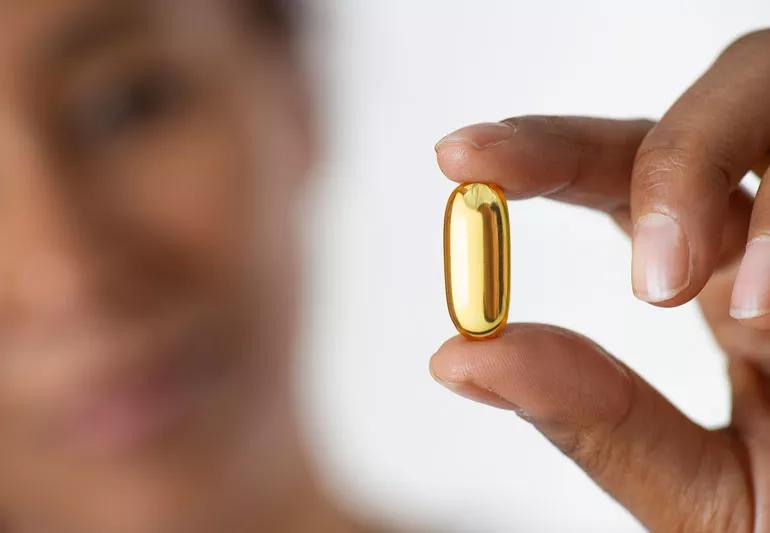
Vitamin D supplementation is common in the United States, with about one-fifth of the adult population taking a daily supplement in one form or another. Although the detrimental effects of insufficient sun exposure in childhood was established centuries ago, the beneficial effects of vitamin D sufficiency have only recently been established, given the myriad investigations associating vitamin D deficiency with numerous chronic diseases. But it is far less clear precisely how to replete low 25-hydroxyvitamin D (25[OH]D) levels, how long treatment should be continued if there are potential hazards in doing so, and how to assess and counsel patients regarding the use of vitamin D. Part one of this review provided a brief historical review, examined how to assess and counsel patients on the use of vitamin D and presented one scenario that clinicians are likely to encounter. Part two offers some more common scenarios and reviews the literature on recommendations for vitamin D supplementation.
An otherwise healthy 30-year-old woman with a BMI of 37 kg/m2 was referred for vitamin D deficiency “unresponsive to D repletion.” Her initial 25(OH)D level was 14 ng/mL. After taking vitamin D2 at a dose of 50,000 IU once weekly with her morning coffee for 4 weeks, her 25(OH)D level remained at 21 ng/mL, still below the normal range.
Advertisement
The clinical challenges with this patient are to consider whether vitamin D2 (ergocalciferol) or D3 (cholecalciferol) makes a difference and whether taking it on an empty stomach is optimal for absorption.
Several recent articles have addressed the question of whether D2 and D3 supplements are equivalent in raising serum 25(OH)D. Houghton and Vieth questioned assumptions about their equivalency and proposed several mechanisms that may contribute to the ability of vitamin D3 to maintain higher serum concentrations over time, including the following:
In a systematic review and meta-analysis, Tripkovic et al concluded that supplementation with vitamin D3 had a significant and positive effect on the raising of serum 25(OH)D concentrations compared with the effect of D2 (P = .001). In a study that explored the relative potency of vitamin D2 and vitamin D3, Armas et al found the 2 forms to be equivalent in absorption. Further, they both produced similar increases in serum 25(OH)D in the first 72 hours, but the 25(OH)D level continued to rise in the D3-treated patients, peaking at day 14. Their calculated area under the curve at 28 days indicated that D3 was 9.5 times more potent than D2.
Does it matter if the supplement is taken on an empty stomach vs with a meal? In a small study, Mulligan and Licata found that taking either vitamin D2 or D3 with the largest meal of the day increased the average serum 25(OH)D level by 50.2% (± 13.4%).
Advertisement
Similarly, a systematic review by Silva and Furlanetto included randomized controlled trials examining the response to a single dose of vitamin D taken with a fat-free meal vs meals that contained 15 g or more of fat. Mean serum 25(OH)D concentrations were higher in those who took the supplement with a meal that included at least 15 g of fat.
This 30-year-old female patient, deemed unresponsive to vitamin D repletion, was treated with vitamin D3 50,000 IU weekly for eight weeks taken with dinner. Her 25(OH)D level rose to 42.8 ng/mL.
A 38-year-old man with a history of fistulizing Crohn’s disease had undergone multiple small-bowel resections and had become dependent on parenteral nutrition. His 25(OH)D level was 12 ng/mL despite taking vitamin D2 50,000 IU daily. In an effort to overcome his malabsorption issue, he would bite into the gel cap to release the contents before swallowing the supplement.
Dual x-ray absorptiometry was notable for an extraordinarily low hip Z-score of −3.4, his long bones were painful to palpation, and his parathyroid hormone level was significantly elevated at 248 pg/mL (reference range 15–65 pg/mL). Osteomalacia is not uncommon in this patient population, but treating the vitamin D deficiency can be very challenging.
In addition to Crohn disease, other conditions can interfere with vitamin D absorption, including a history of malabsorptive-type bariatric surgery, celiac disease, cystic fibrosis, steatorrhea, short bowel disease, inflammatory bowel disease, and severe cholestasis. A vitamin D challenge test is one way to confirm the absorptive capability for vitamin D supplementation in these patients.
Advertisement
When vitamin D is taken orally, it is incorporated into the chylomicron fraction, and about 80% of the dose is absorbed into the lymphatics. The blood level of 25(OH)D will peak about 12 hours after a single dose of 50,000 IU. Knowing this about oral absorption of vitamin D allows for provocative testing in patients with suspected malabsorption of the vitamin.
To test for malabsorption, a blood sample is drawn immediately before administering a 50,000-IU oral dose of vitamin D. The blood draw is repeated in 12 to 24 hours. If no increase in 25(OH)D is noted, the patient has “complete” malabsorption of vitamin D. Incidentally, if this is the finding, then the patient may need testing for deficiencies of other fat-soluble vitamins such as vitamin A.
In addition to supplementation, vitamin D synthesis can take place when the skin is exposed to UV-B light. The therapeutic benefits of phototherapy are recognized for a wide variety of skin conditions, and with careful skin-typing and carefully metered exposure to UV-B light, phototherapy can also achieve normal 25(OH)D levels.
This 38-year-old patient was referred to dermatology for phototherapy. UV-B light was administered three days per week under the close supervision of an experienced dermatologist, and his 25(OH)D level rose to 48 ng/mL within a few weeks.
Sunbathing and tanning booths are not therapies for vitamin D deficiency. Sunshine is composed of approximately 95% UV-A and 5% UV-B, but only UV-B is required for vitamin D synthesis. UV-A is the predominant or sole light source used in tanning beds, and the dose of UV-A in tanning beds can be up to 12 times that provided by the sun.
Advertisement
Skin cancers comprise one-half of all cancers, and UV-A and UV-B are both implicated. UV-A is thought to damage skin and increase the risk of melanoma by causing oxidative stress-induced DNA damage. UV-B damage is more direct, with photoproducts that are implicated in skin carcinogenesis. Skin type and age are factors in the response to UV exposure, but in general, exposing 5% of the body surface twice weekly for 20 minutes during the summer months may be equivalent to 430 IU of vitamin D per day, with a plateau being reached after 20 minutes.
A 78-year-old otherwise healthy woman with primary hyperparathyroidism also has vitamin D deficiency, with a 25(OH)D level of 15 ng/mL in the presence of an elevated serum calcium level of 11.4 mg/dL (reference range 8.5–10.2 mg/dL), high parathyroid hormone of 128 pg/mL (reference range 15–65 pg/mL), low phosphorus of 1.7 mg/dL (reference range 3.0–4.5 mg/dL), and high 24-hour urine calcium of 472 mg (reference range 100–300/day).
In a meta-analysis of 10 studies that included 340 patients with primary hyperparathyroidism, Shah et al assessed the effect of 25(OH)D replacement in patients with coexistent vitamin D deficiency. The studies included the use of vitamin D2 and D3 supplements, and the time span of administration ranged from one to 12 months. Interestingly, this study noted a nonsignificant but modest decline in serum calcium after vitamin D replacement. Only 2.2% developed more severe hypercalcemia (> 12 mg/dL) that responded to stopping the supplement or to reducing the dose. The authors concluded that vitamin D replacement in patients with primary hyperparathyroidism does not worsen hypercalcemia.
This patient was placed on 5,000 IU of vitamin D3 daily, taken with her largest meal, and was maintained on that dose following parathyroid surgery. At her 3-month postoperative visit, the 25(OH)D level was normal at 52 ng/mL, and her parathyroid hormone and serum calcium levels were also normal.
Vitamin D toxicity can result from overcorrection of vitamin D deficiency. Case reports have implicated manufacturing errors, overdosing by patients or prescribers, or a combination of these factors. Perhaps no report is more poignant than the report by Zhou et al of an 80-year-old man who presented with signs and symptoms consistent with vitamin D toxicity including confusion, dysarthria, and ataxic gait, and was found to have a serum calcium of 14.4 mg/dL in the presence of a parathyroid hormone level of 11 pg/mL and 25(OH)D of 365 ng/mL. He had been prescribed a weekly 50,000-IU vitamin D tablet, but at some point, he began to take it daily with his other medications. All of his symptoms resolved after a brief hospital stay, during which the vitamin D supplement was stopped and the hypercalcemia was addressed.
Vitamin D deficiency is relatively common. The detrimental effects of vitamin D deficiency have been well documented, dating to the 1600s, but only during the early 1900s did we discover and implement palatable fortification of milk and other foods that led to the eradication of rickets in children. However, fortification of milk alone failed to eliminate vitamin D deficiency.
Fortunately, vitamin D supplements are easily prescribed, inexpensive, and available over the counter. It is important for clinicians to be attentive to the likelihood of vitamin D deficiency, especially in patients with certain diseases and conditions; to advise patients on the best ways to attain and maintain an adequate 25(OH)D level; to counsel patients taking supplements on avoiding oversupplementation; to advise against inappropriate reliance on sun exposure and tanning beds for vitamin D supplementation; and to recognize symptomatic vitamin D toxicity.
Advertisement

Advocacy group underscores need for multidisciplinary expertise

A reconcilable divorce

A review of the latest evidence about purported side effects

High-volume surgery center can make a difference

Advancements in equipment and technology drive the use of HCL therapy for pregnant women with T1D

Patients spent less time in the hospital and no tumors were missed

A new study shows that an AI-enabled bundled system of sensors and coaching reduced A1C with fewer medications

Association revises criteria for the diagnosis and resolution of severe conditions