Locations:

A proof-of-concept study shows how 3D pouchography can be a useful adjunct for surgeons
Image content: This image is available to view online.
View image online (https://assets.clevelandclinic.org/transform/778d581a-747d-4b8c-93fa-2a4873517b8f/Holubar_-j-pouch)
Stefan Holubar speaking with researcher
Currently, surgeons involved with ileal pouch-anal anastomosis (IPAA) construction rely on two-dimensional (2D) fluoroscopic and cross-sectional imaging to diagnose pouch complications and plan pouch salvage surgery. However, there are limitations involved with 2D visualization since surgeons are only able to see parts of the pouch and pouch staple lines on any given 2D image cross-section.
Advertisement
Cleveland Clinic is a non-profit academic medical center. Advertising on our site helps support our mission. We do not endorse non-Cleveland Clinic products or services. Policy
A recent study appearing in the Journal of Crohn’s and Colitis describes a proof-of-concept study of 3-dimensional (3D) pouchography using virtual and printed 3D models of ileal pouch-anal anastomosis. The research group performed a retrospective, descriptive case series of a convenience sample of 10 pouch patients with or without pouch dysfunction who had CT scans appropriate for segmentation. The group believes that 3D technology can be used to visualize healthy and dysfunctional pouches and assist with the diagnosis of pathologies that result in pouch dysfunction.
“There have been several technological advances associated with IPAA construction, including laparoscopic, robotic and transanal techniques,” says the study’s corresponding author Stefan Holubar, MD, of the IBD Surgery Section and Director of Research, Department of Colorectal Surgery, Cleveland Clinic. “But the complex 3D anatomy of IPAAs can lead to a variety of structural anomalies. We felt that 3D pouchography could provide surgeons with a more comprehensive understanding of normal and pathologic pouch anatomy, which could in turn help reduce instances of pouch dysfunction.”
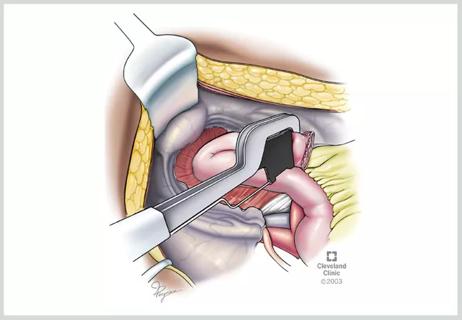
Using the Cleveland Clinic Pouch Registry, the research group identified a convenience sample of 10 patients with IPAA who had a diagnosis of ulcerative colitis or Crohn’s disease and who had computed tomography (CT) scans appropriate for segmentation. The patient’s CT scans were uploaded to a software program that manipulated system-generated 3D reconstructed images. These images included only bones, vasculature, surgical staples and hardware. The bone features were then removed, and vasculature and anatomic features other than pouch staples were also excluded. The pouch staples were then centered and zoomed, and the contrast was adjusted to increase their intensity. The resultant 3D models could be rotated along the vertical (anterior-posterior), lateral (left-to-right) and longitudinal (craniocaudal or head-foot) axes. These images were available to clinicians caring for the patients, and representative still images and cine clips were also made available for annotation.
Advertisement
“The second part of our study involved additional manual segmentation of the 3D models,” says Dr. Holubar. “These were done by one of the other researchers on our team, Nour Mohammed, MS.”
Dr. Mohammed was able to augment the segmented models for educational purposes by adding voxels to fill in gaps in the staple lines to make them continuous and suitable for 3D printing. A semi-automated, region-of-interest capture tool was also used to add to the bowel wall. These 3D virtual models were then exported to 3D PDF files for annotation and 3D printing of deformable models with a translucent bowel wall and opaque staple lines.
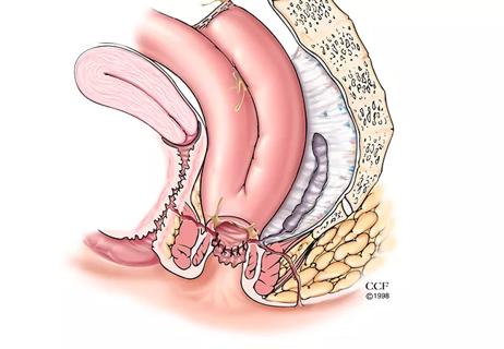
“We observed that the 3D pouch reconstruction reliably demonstrated multiple features based on the staple lines alone,” says Dr. Holubar. “This includes the pouch body staple lines, tip of the J staple line, and anorectal transverse and circular staple lines. We also found that the 3D modeling can reveal the relationship between these structures. For example, if the tip of the J was constructed flush with the pouch, we noticed that it appeared contiguous with the pouch body staple lines. However, in instances where the tip of the J is 1 or 2 cm long, the transverse staple line appears separate— almost ‘floating’—next to the pouch body. Essentially, the tip of the J location on the model can be used as a reference point.”
Dr. Holubar notes the team was able to determine the relationship and variations between the anorectal transverse and circular staple lines. They found that if the spike was placed through the center of the transverse staple line, the resultant dog ears were visible. On the other hand, if the spike was placed through one corner, a tennis racquet shape was visible instead.
Advertisement
The pouch’s shape and orientation were also evident in pouch 3D reconstructions. The research group observed several variations including straight tubular, “C” shaped and reverse “C” shaped, and spiraled. In terms of the pouch orientation, the group noticed that while the anterior pouch-body linear staple line should be the anterior midline, the pouch can shift and get displaced to one side or the other. In this patient cohort, the group observed cases in which the pouch was rotated to the left, but not more than 90 degrees, in normal pouches.
Video content: This video is available to watch online.
View video online (https://cdnapisec.kaltura.com/p/2207941/sp/220794100/playManifest/entryId/1_mwrauxj9/flavorId/1_5f3sgelj/format/url/protocol/https/a.mp4)
3D model of a perfect pouch with zero rotation. The model clearly shows the spur with lack of staples posterior as it should be. The bright spots are the "jags" from the serial firing of the GIA-100 stapler, and the short tip of J is on the right. The model also shows the circular staple line at the bottom with short dog ears.
“At Cleveland Clinic, we have been incorporating 3D pouch segmentation into our care,” explains Dr. Holubar. “It’s allowed us to better understand some of the mechanical pathologies with pouch dysfunction that we come across when caring for patients, so we can plan pouch salvage surgery and relieve patients' often severe symptoms, improve their quality of life and save their pouch.”
The research paper describes three different scenarios of pouch dysfunction in which 3D pouch segmentation has helped the team better gain insight. For patients with pouch septum, the researchers observed an enterolith formation and noted that pouch strictures related to Crohn’s-like disease of the pouch are not uncommon, and these can result in proximal pouch body dilation and a dilated, heart-shaped, proximal pouch.
In cases of acute pouch volvulus, spiraling staple lines may be observed; and in cases of chronic pouch volvulus that result in pouch dysfunction, spiraling of the pouch body staple lines can be observed endoscopically and radiographically.
Advertisement
“We found that 3D pouch reconstruction was particularly effective in cases of twisted pouch syndrome (TPS),” says Dr. Holubar. “TPS is one of the more difficult conditions to diagnose preoperatively, and actually, the impetus for developing this study was based on the need to be able to visualize the entire pouch body staple line.”
He notes that TPS typically results from one of three reasons: spiraling of the distal pouch body during firing of the circular stapler, adhesions from the occult tip of J leaks, or unintentional pouch construction in a 180° manner with the mesentery anterior or a full 360° from a twisted mesentery. In cases of the latter two reasons, the model was particularly effective. In cases of unintentional 180°, a clinician might see the spur with a lack of staples to be anterior instead of posterior, and in cases of unintentional 360°, the distal portion of the superior mesenteric artery (SMA) spiraled 360°. However, following redo IPAA, a normal pouch can be seen on the 3D model.
“3D pouchography has been an extremely useful tool for managing patient care and improving how we plan pouch salvages,” says Dr. Holubar. “It provides us with such a comprehensive understanding of each patient. Being able to annotate these models and share notes with other providers also improves our overall communication so that we can ensure everyone is on the same page. We believe that with the rapid developments, we’re seeing in the field of artificial intelligence machine vision algorithms, we’ll be able to improve these models and make them even more accurate and nuanced. Presently, we are implementing a blinded study to assess the value of this technique in the diagnosis of mechanical pouch complications.”
Advertisement
Advertisement

Retrospective analysis looks at data from more than 5000 patients across 40 years
Three presentations from the DDW 2023 conference help identify best candidates and illustrate the positive impact redo IPAAs can have on a patient’s QoL

Radiation to the pelvis from cancer treatment made the traditional treatment path unavailable
A new study illustrates why it’s important for clinicians to be aware and suspicious of tip of the J-pouch leaks

Landmark trial data suggest appendectomy may reduce relapse and improve remission rates in select patients

Implications for surgical decision-making and patient management

Dr. Prabhu discusses mentorship, collaboration and her vision for the future of the department

New program looks to innovative approaches for advancing early detection and multicancer prevention