A glimpse into our centralized monitoring strategy and more

A few years ago, Cleveland Clinic instituted a novel concept — an off-site unit providing round-the-clock cardiac telemetry monitoring for non-critically ill patients in Cleveland Clinic hospitals — under the direction of electrophysiologist Daniel Cantillon, MD. His colleague, Steven Nissen, MD, Chief Academic Officer of Cleveland Clinic’s Miller Family Heart & Vascular Institute, recently caught up with Dr. Cantillon for an update on this and other advanced approaches to monitoring. An edited transcript of their conversation follows.
Advertisement
Cleveland Clinic is a non-profit academic medical center. Advertising on our site helps support our mission. We do not endorse non-Cleveland Clinic products or services. Policy
Dr. Nissen: Can you give us an overview of how the central monitoring unit works and why it’s significant?
Dr. Cantillon: The central monitoring unit is an off-site facility that provides secondary monitoring for hospitalized patients in non-intensive care units on Cleveland Clinic’s main campus and in 10 Cleveland Clinic regional hospitals. The monitoring is provided 24/7 through a series of innovative technologies and methods developed in-house to get out in front of decompensating patients by enabling advanced detection of the changes associated with decompensation.
Dr. Nissen: So the unit is remotely located. I think a lot of physicians might find it surprising that the folks doing the monitoring are not actually here in the hospital.
Dr. Cantillon: It’s our command center. It’s a place where we take an extraordinary workforce of trained technicians and remove them from the distractions of hospital activities and noises so they can focus on identifying those patients who are at risk of decompensation. They do it with the support of our technologies and risk-stratification tools, with the goal of getting ahead of cases of cardiac arrest, which of course is the key to improving survival outcomes in resuscitation.
Dr. Nissen: How many people are monitored on a given day?
Dr. Cantillon: We’re close to a capacity of 2,000 patients per day. Of that, we run a census between 1,100 and 1,200 patients depending on bed occupancy rates across the hospitals.
Dr. Nissen: How many people are sitting in front of a monitor at any given time?
Advertisement
Dr. Cantillon: We have a workforce of about a dozen technicians who provide direct coverage of the central monitoring station at a given time. We also have lead technicians who serve in an oversight capacity.
Dr. Nissen: How do a dozen people look after more than a thousand patients? That’s extraordinary.
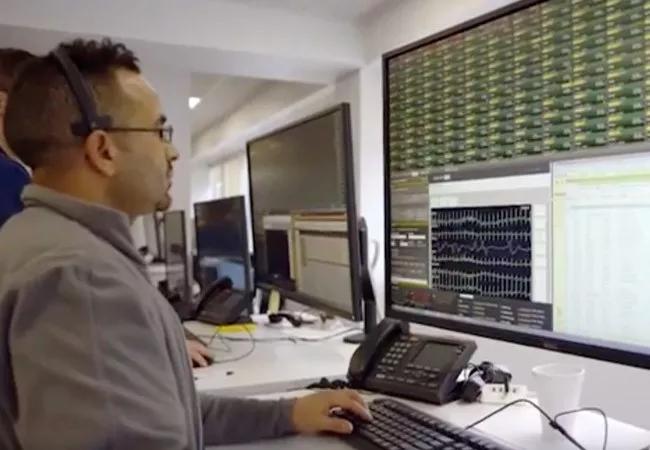
Dr. Cantillon: That’s where the technology comes in. We developed, validated and deployed a clinical algorithm that serves to bring to the attention of the technicians only those few patients — from among the thousand-plus patients being monitored — who may require closer attention at a given time. That closer attention involves looking at the patient’s chart, their ECG waveform, their vital sign trends, etc., on a computer monitor to understand what’s happening with the patient [see photo below]. The technician then makes a well-informed phone call to the bedside nursing team and occasionally, on a discretionary basis, directly to our emergency response teams.

Image content: This image is available to view online.
View image online (https://assets.clevelandclinic.org/transform/8d3e0db0-c433-4d2f-bcfa-e055fb5b49b1/19-HRT-3750-central-monitoring-unit-inset_jpg)
Dr. Nissen: Exactly what’s being monitored by the central unit?
Dr. Cantillon: There are three components to our algorithm. First is the reason the patient was put on the monitor. We essentially determine the patient’s pre-test probability of having a cardiac event. For example, was the patient transferred from another hospital for coronary revascularization or were they admitted for syncope? We know the specific event rate for each category like that, and it’s factored into the algorithm.
The second component involves changes in the patient’s vital signs — heart rate, blood pressure, respirations, pulse oximetry — with patients serving as their own controls. So we look at changes occurring in real time compared with the patient’s own moving average, and we pick up on variations that signal the patient is moving in the wrong direction. The algorithm scores the variations and puts them into the equation.
Advertisement
The third component involves the nominal alarms that come from our vendors, such as telemetry systems and premature ventricular contraction alarms. We’ve found that most of those are not clinically meaningful, but we’ve reclassified each of those alarm codes and assigned them a value based on a review of the clinical evidence.
So the three components are combined to generate a total priority risk score for the patient. When that score reaches a certain threshold, the patient’s tile on the monitor turns red. That prompts the technician to open the patient’s tile for a detailed view showing information such as their live ECG waveform and vital sign trending data. It also opens our EMR system to show laboratory test results. The technician is able to dive into the context of why the algorithm triggered the alert.
Dr. Nissen: That’s in contrast to the traditional practice of waiting until a patient has ventricular tachycardia and alarms are suddenly going off and everyone’s running around.
Dr. Cantillon: At which point it’s too late. We know that in those circumstances where we wait until ventricular tachycardia arrest occurs, we are not very successful in resuscitating the patient. But if we get out in front of it within an hour before that, we know, based on the outcomes we published in JAMA (2016;316:519-524), that we can achieve up to 93% return of spontaneous circulation. In the world of hospital medicine, that’s unprecedented.
Dr. Nissen: How accurate is the system? If a patient’s tile on the monitor turns red, what’s the probability that there’s really something bad going on versus it being a false alarm?
Advertisement
Dr. Cantillon: It’s quite high. We find that up to a third or even half of patients whose tiles turn red actually have an actionable event that requires the bedside team to do something different, such as change the patient’s level of care, make a medication adjustment or order some tests. Conversely, we’ve found that over 99% of the alarms in traditional alarm systems are inactionable. They’re actually contributing to background noise that can drown out the signal of the patient who truly requires our attention. It’s classic alarm fatigue that undermines the whole purpose of alarms in the first place.
Dr. Nissen: You mentioned publishing on the results of this approach. Tell us about the key findings.
Dr. Cantillon: The main finding, as reported in JAMA, was that in the situations where the central monitoring unit allowed us to get out in front of these events and our emergency response teams were deployed, we were able to achieve a 93% return of spontaneous circulation. American Heart Association statistics show that 1 in 10 patients survive a cardiac arrest in the community, and 1 in 4 in a non-ICU hospital setting. So our results represent a paradigm shift in what can be achieved.
And as we get better and smarter about advanced cardiac waveform analytics and really get into the embedded signals within the ECG waveform, we believe we can achieve even better outcomes. Right now we identify decompensating patients about 80% of the time, so that means we’re not getting out in front of the event about 20% of the time. We see this as an opportunity to dive in and try to understand what subtle signals we may be missing. We’re also exploring how machine learning techniques and technologies might help identify those subtle patterns.
Advertisement
Dr. Nissen: What about something like ST depression? Is that yet part of your algorithm?
Dr. Cantillon: It will become part of it. We’re looking at wavelet-type recognition. Much of our work in this space has been inspired by the way pacemakers and defibrillators can discriminate different types of arrhythmias. Many of these devices work by using wavelet recognition: They take a wavelet of a patient’s normal rhythm, they freeze that as a template and they compare it to dynamic changes occurring in real time. We believe that same phenomenon can occur for the ECG waveform. So we’re looking at subtle changes with ST depression, ST elevation, QT prolongation — potential warning signs that the patient is having a meaningful change in clinical status but that don’t register on a nominal vendor alarm system.
Dr. Nissen: That’s amazing. One last question: What if a technician calls from the remote unit and nobody answers the phone?
Dr. Cantillon: We have an extensive escalation protocol where the call then goes to the charge nurse, and then to the nurse manager. The entire approach of the central monitoring unit — from the methodology to the workflows to the software to the training programs for our technicians — has been designed and built in-house in a structured and interconnected way. It’s been an extraordinary privilege to be at the center of an undertaking of this scale and potential significance.
This Q&A was derived from an episode of Cleveland Clinic’s “Cardiac Consult” podcast for healthcare professionals. To hear the 15-minute discussion between Drs. Cantillon and Nissen, listen or subscribe from your favorite podcast source.
Advertisement

How Cleveland Clinic is using and testing TMVR systems and approaches

NIH-funded comparative trial will complete enrollment soon

How Cleveland Clinic is helping shape the evolution of M-TEER for secondary and primary MR

Optimal management requires an experienced center

Safety and efficacy are comparable to open repair across 2,600+ cases at Cleveland Clinic

Why and how Cleveland Clinic achieves repair in 99% of patients

Multimodal evaluations reveal more anatomic details to inform treatment

Insights on ex vivo lung perfusion, dual-organ transplant, cardiac comorbidities and more