Study of bone bruise patterns indicates more medial damage than expected

Knee rotation may not be the primary cause of anterior cruciate ligament (ACL) ruptures after all. For decades, orthopaedic providers have explained ACL tears as resulting from a severe twist in the knee joint, a belief reinforced by the presence of bruising on the lateral femoral condyle and lateral tibial plateau. These lateral bone bruises, visible on MRI, are prevalent in patients with a torn ACL.
Advertisement
Cleveland Clinic is a non-profit academic medical center. Advertising on our site helps support our mission. We do not endorse non-Cleveland Clinic products or services. Policy
A recent study led by Cleveland Clinic has shown that bone bruising also occurs in the knee’s medial compartment after an ACL tear. And the position of the medial bruises indicates a completely different mechanism of injury.
“The concept was suggested at an Orthopaedic Research Society meeting a few years ago,” says sports medicine orthopaedic surgeon Kurt Spindler, MD, Director of Clinical Research and Outcomes at Cleveland Clinic Florida. “A researcher proposed that it’s not really a rotational load or torque but a straight anterior load or force that causes most ACL tears. They pointed to a limited amount of data showing a high prevalence of medial bone bruises. The concept wasn’t taken all that seriously at the meeting, but our research team decided to explore it using our own data. Sure enough, that initial researcher was right.”
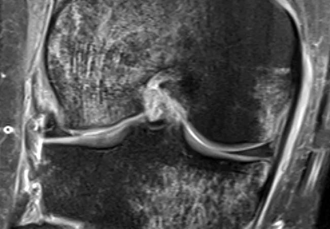
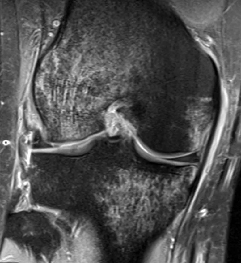
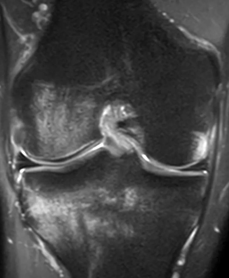
In the Cleveland Clinic-led study published in the Orthopaedic Journal of Sports Medicine, researchers studied the MRI scans of more than 200 patients who had ACL reconstruction between 2015 and 2017. More than 80% of the patients had bone bruises in the medial compartment or both medial and lateral compartments.

Image content: This image is available to view online.
View image online (https://assets.clevelandclinic.org/transform/74dd9064-e5d2-406d-8f6f-aa24d8cdd8c7/22-ORI-3418208-CQD-Inset1-jpg)
Coronal MRI showing bruising to medial tibial plateau and medial and lateral femoral condyles one month after ACL rupture in a 28-year-old patient.

Image content: This image is available to view online.
View image online (https://assets.clevelandclinic.org/transform/969de18e-c176-4fba-9c6b-8905a110ca1f/22-ORI-3418208-CQD-Inset2-jpg)
Coronal MRI showing bruising to medial femoral condyle three weeks after ACL rupture in a 27-year-old patient.

Image content: This image is available to view online.
View image online (https://assets.clevelandclinic.org/transform/65e98933-e9f6-4681-97cb-fc420de9bc01/22-ORI-3418208-CQD-Inset3-jpg)
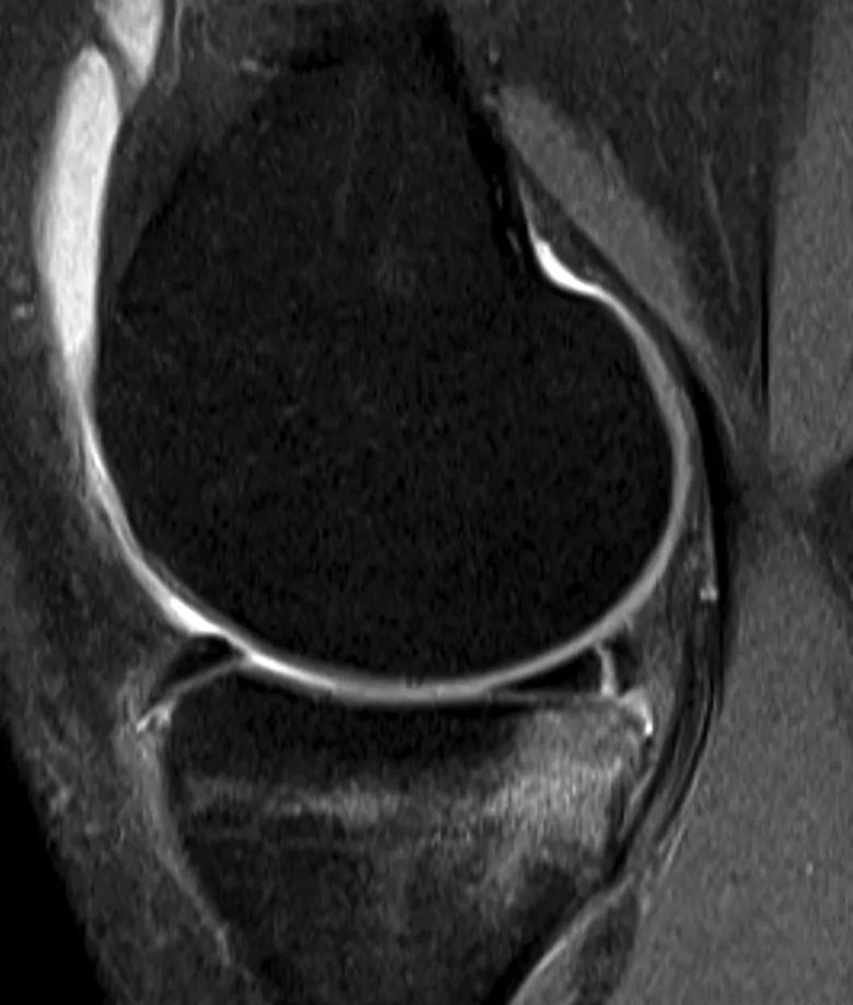
Sagittal MRI showing bruising to medial tibial plateau two weeks after ACL rupture in a 24-year-old patient.
Nearly all bruises (98%) in the medial tibial plateau were in the posterior third of the plateau. This is an important detail because it suggests that anterior tibial translation — and not hyperextension and rotation — contributes to an ACL rupture.
“Bone bruises on the back of the tibial plateau and the front of the femoral condyle indicate the occurrence of an anterior subluxation of the tibia on the femur,” explains Dr. Spindler. “And because the majority of patients have bruising on the medial side, the mechanism of injury is more likely a straight anterior pull rather than a rotation.”
Advertisement
More medial compartment damage occurs with a torn ACL than previously thought, he adds. For the anterior femur to bruise the posterior medial tibial plateau, it must first contact the posterior horn of the medial meniscus.
“That posterior horn is undergoing some damage that we didn’t recognize before,” says Dr. Spindler. “The posterior horn of the medial meniscus is an important protector of articular cartilage in the knee. Damage to the meniscus could lead to post-traumatic arthritis, which would have a significant negative effect on knee function.”
A more complete understanding of the mechanism of injury could improve prevention strategies and treatments.
“When preparing for ACL reconstruction, surgeons should look more carefully at the medial side of the knee to assess damage there, especially damage to the posterior horn of the medial meniscus,” says Dr. Spindler. “That will inform a more complete evaluation and treatment for an ACL-injured knee.”
Redesigned knee braces or rehabilitation strategies to guard against ACL rupture, including repeat rupture after ACL reconstruction, should aim to prevent anterior subluxation of the tibia in addition to rotation, he says.
Advertisement
Advertisement

Nonthermal technique reduces bleeding and perforation risk

Standardizing a minimally invasive approach for Barrett’s Esophagus and Esophageal Cancer

PSMA-targeted therapy for metastatic prostate cancer now offered at Cleveland Clinic Weston Hospital

Nationally recognized urologic oncologist offers vision for growth, innovation, and excellence

Noninvasive modality gains ground in United States for patients with early-to-moderate disease

Cleveland Clinic Weston Hospital’s collaborative model elevates care for complex lung diseases

Interventional pulmonologists at Cleveland Clinic Indian River Hospital use robotic technology to reach small peripheral lung nodules

Trained in the use of multiple focal therapies for prostate cancer, Dr. Jamil Syed recommends HIFU for certain patients with intermediate-risk prostate cancer, especially individuals with small, well-defined tumors localized to the lateral and posterior regions of the gland.