Locations:

Full interpretation changes focus of case
In this video, Sanjay Mukhopadhyay, MD, director of pulmonary pathology in the Department of Anatomic Pathology, presents an unusual case of interstitial lung disease in which the focus on atypical epithelial cells changed completely once a full pathology interpretation was performed. The referring pathologist suspected adenocarcinoma; Dr. Mukhopadhyay explains why another diagnosis is a better fit below.
Advertisement
Cleveland Clinic is a non-profit academic medical center. Advertising on our site helps support our mission. We do not endorse non-Cleveland Clinic products or services. Policy
Video content: This video is available to watch online.
View video online (https://www.youtube.com/embed/ELnYD2tUmT4?feature=oembed)
Pathology Insights: Atypical Epithelial Cells with Sanjay Mukhopadhyay, MD
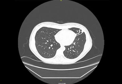
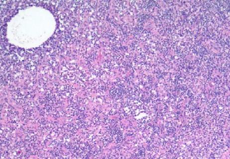
Dr. Mukhopadhyay describes a resection specimen of a fibrotic nodular lesion with atypical epithelial cells from a patient with emphysema and a history of smoking evidenced by the presence of smoking-related interstitial fibrosis. The key diagnostic finding in this case lies in a tiny, nodular stellate scar. A mix of plasma cells, lymphocytes, chronic inflammatory cells, eosinophils and a few histiocyte-like cells clue Dr. Mukhopadhyay into a diagnosis of Langerhans cell histiocytosis. CD1a and S-100 immunostaining confirmed the presence Langerhans cells.
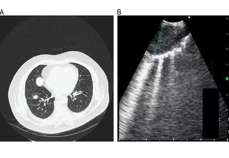
Dr. Mukhopadhyay returns to the original lesion of concern to demonstrate typical features of the scarred phase of Langerhans cell histiocytosis. “Langerhans cell histiocytosis in the lung is seen nearly exclusively in cigarette smokers,” he notes, “and in these patients you get nodular lesions in addition to SIRF and respiratory bronchiolitis in the background lung. You get these tiny nodules or scars that are filled with Langerhans cells and with other inflammatory cells.”
Pulmonary pathology at Cleveland Clinic
Cleveland Clinic houses one of the few completely subspecialized thoracic pathology services in the country that handles large volumes of neoplastic and non-neoplastic lung diseases. The Pulmonary Pathology Section receives approximately 5,000 in-house specimens a year, including core needle biopsies, bronchoscopic biopsies, transbronchial cryobiopsies, surgical lung biopsies for interstitial lung disease, transplant transbronchial biopsies and pleural and mediastinal biopsies. Larger specimens include surgical resections for lung cancer, primary metastatic neoplasms, thymic neoplasms, mesothelioma and a large volume of explanted lungs.
Advertisement
Dr. Mukhopadhyay’s team utilizes state-of-the-art molecular testing, including a comprehensive lung Next-Generation Sequencing (NGS) panel and FISH for ALK, ROS1 and RET, as well as a large panel of immunohistochemical and molecular tests to diagnose and classify lung carcinoma, mesothelioma, mediastinal pathology, interstitial lung disease and a variety of non-neoplastic lung diseases.
Advertisement
Advertisement

Screen patients seeking care for chlamydia, gonorrhea
Lingulectomy removes infection when antibiotics fail
Researchers have developed immunoprofiles for an emerging disease with a mortality rate as high as 27%

Findings from one of the first published case series
Don't discount this crucial step
EBUS-TBNA found safe and effective
How to spot the rare infection
Not all lung nodules are malignant