Locations:

Normal tissue or thymoma?
In this video, Sanjay Mukhopadhyay, MD, staff thoracic pathologist in the Department of Anatomic Pathology, presents a case that demonstrates the differences in thymic architecture that indicate thymoma versus normal thymic tissue. He also differentiates between Type A and Type B thymomas. Get the basics in this brief introduction:
Advertisement
Cleveland Clinic is a non-profit academic medical center. Advertising on our site helps support our mission. We do not endorse non-Cleveland Clinic products or services. Policy
Video content: This video is available to watch online.
View video online (https://www.youtube.com/embed/aqIbLduZH20?feature=oembed)
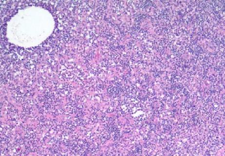
The case Dr. Mukhopadhyay discusses involves thymic pathology, which many pathologists see very seldom. He shows normal thymic tissue contrasted with abnormal tissue. Normal thymic tissue has a lot of adipose tissue in the background, and thymic tissue is sprinkled in, appearing as little blue areas in the slide. The blue areas contain lighter and darker areas that contain both epithelial and lymphoid cells in the thymic tissue, and these cells are typically rich in TdT-positive lymphocytes, immature lymphocytes that occur in the normal thymus. The TdT-positive lymphocytes are mixed with epithelial cells that are slightly larger and sprinkled together with adipose tissue.
Dr. Mukhopadhyay also identifies Hassall’s corpuscles, epithelial cells that appear vaguely squamoid and are scattered within the thymic tissue. Normal thymic architecture, then, appears as little nests of thymic tissue including epithelial and lymphoid cells, sprinkled among adipose tissue, with occasional Hassall’s corpuscles among the tissue.
Abnormal thymic architecture, on the other hand, looks entirely different from slides of normal architecture. The nests appear vaguely thymic but are much bigger and broader and have completely obliterated the normal architecture. The nests are separated by pink bands of fibrous tissue. This appearance is the classic low-power architecture of thymoma.
This video is part of the Pathology Insights series, featuring important cases, methods and practices personally presented by staff pathologists. The short videos break down information about interesting pathology cases to better inform doctors, laboratory staff, patients or anyone interested in the field of pathology.
Advertisement
Advertisement

Screen patients seeking care for chlamydia, gonorrhea
Lingulectomy removes infection when antibiotics fail
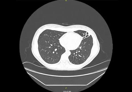
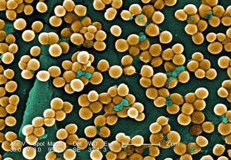
Researchers have developed immunoprofiles for an emerging disease with a mortality rate as high as 27%

Findings from one of the first published case series
Don't discount this crucial step
EBUS-TBNA found safe and effective
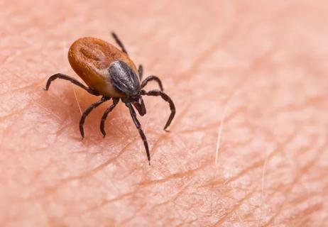
How to spot the rare infection
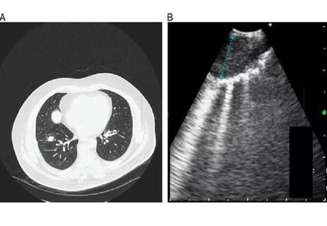
Not all lung nodules are malignant