Locations:

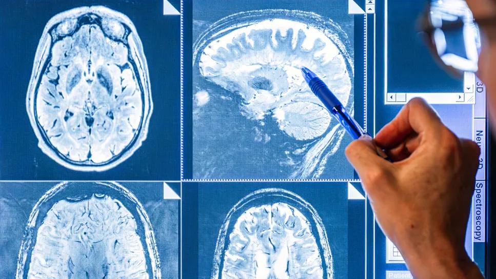
Image content: This image is available to view online.
View image online (https://assets.clevelandclinic.org/transform/cfa0e190-ce45-4367-9de0-3968b92116f3/hercules-study-multiple-sclerosis)
Brain scans
While stroke may get the lion’s share of public awareness and research funding among cerebrovascular diseases, diagnostic and treatment advancements are providing physicians better options for helping patients avoid the devastating consequences of many chronic conditions that affect blood flow and the blood vessels in the brain.
Advertisement
Cleveland Clinic is a non-profit academic medical center. Advertising on our site helps support our mission. We do not endorse non-Cleveland Clinic products or services. Policy
“It’s gratifying to see the progress we’ve made in caring for patients with chronic cerebrovascular conditions, even in just the past decade since completing my endovascular neurosurgery fellowship,” says Michal Obrzut, MD, Head of the Section of Neurointerventional Radiology at Cleveland Clinic Weston Hospital.
Dr. Obrzut is part of a team of specialists with the Neurosciences Institute at Cleveland Clinic in Florida that has developed new imaging protocols and adapted emerging and innovative endovascular treatments to care for patients with chronic subdural hematomas (cSDH), cerebral aneurysms, and spinal dural arteriovenous fistulas (sDAVF).
cSDH is an increasingly common neurological condition associated with older adults who experience head trauma. Often slow to develop, it is caused by an initial venous bleed that coagulates and forms a clot in the space below the dura mater, between the brain and the skull.
“Just two years ago surgical evacuation was the treatment of choice, but this has a high risk of recurrence,” explains Dr. Obrzut. “The approach is based on the long-held theory that a chronic subdural hematoma is the result of continued bleeding from ruptured bridging veins within the subdural space.”
According to Dr. Obrzut, a better understanding of cSDH pathophysiology embraced over the past decade indicates that hematoma recurrence may be caused by blood leaking from fragile arteries that arise following SDH coagulation and clot formation.
“Neovasculature develops to feed the membrane that forms around the clotted blood, as part of the body’s inflammatory response,” says Dr. Obrzut. “A growing body of research has shown that these small arteries, which are supplied by the middle meningeal artery, are the source of rebleeding.”
Advertisement
This new understanding, though not yet universally accepted, has led to the development of an innovative endovascular treatment for cSDH: middle meningeal artery (MMA) embolization.
The team at Weston Hospital uses an endovascular approach to deliver a liquid embolic injection of n-BCA glue into branches of the MMA to cut off blood supply to the membrane of the hematoma and prevent rebleeding.
“The approach helps tip the balance towards resorption and hematoma resolution,” confirms Dr. Obrzut. “We‘ve successfully treated more than 100 patients with MMA embolization either as a stand‐alone treatment or as an adjunct to surgery.”
Endovascular advancements have had a profound affect in treating another relatively common chronic cerebrovascular condition. Approximately 6.7 million people in the United States are estimated to have an unruptured brain aneurysm, according to the Brain Aneurysm Foundation, and about 30,000 will suffer a ruptured aneurysm each year.
“Fortunately most cerebral aneurysms are small and do not require treatment,” says Dr. Obrzut. “These are usually found incidentally during brain imaging.”
Depending on its type, size and location, an aneurysm may merit a watchful waiting approach to monitor its development. The Weston Hospital team developed an imaging protocol to safely follow asymptomatic patients with unruptured aneurysms that minimizes the use of cerebral angiograms, which have a rare risk of stroke.
“We typically perform a magnetic resonance angiography, using a 3 Tesla MRI, for the initial evaluation, followed six months later with an MRA to determine if the aneurysm is growing or stable,” explains Dr. Obrzut. “Depending on the growth rate observed, the patient may be followed with a yearly MRA.”
Advertisement
The treatment for unruptured aneurysms that require intervention has greatly evolved over the past three decades. Today, the majority of patients are treated endovascularly, with only a small percentage requiring open surgery.
“Our team offers the full gamut of endovascular treatment options, including coil embolization, stent-assisted coil embolization, and flow diversion,” says Dr. Obrzut. He notes Weston Hospital was the first center in South Florida to use a third-generation flow diversion device that incorporates surface modification technology to reduce thromboembolic complications.
“The shield technology acts as a ‘cloaking device’ from platelets, decreasing the occurrence of implant material thrombogenicity,” explains Dr. Obrzut. “This allows us to reduce the use of blood thinners and lowers the risk of stroke.”
For patients who require surgery, the team performs microvascular clipping using advanced intraoperative imaging techniques.
Dr. Obrzut and his colleagues also have been involved in advancing care for patients with spinal dural arteriovenous fistulas (sDAVFs), the most common type of spinal vascular malformation.
A sDAVF develops when a radiculomeningeal artery communicates directly with a radicular vein, causing venous congestion and hypertension to occur. This can lead to hypoxia and ultimately spinal cord dysfunction.
“These malformations are rare, slow to develop, and often misdiagnosed,” says Dr. Obrzut. “They typically occur in older men, who will present with progressive spinal cord symptoms such as paresthesia, gait difficulty, and changes in bowel or bladder function.”
Advertisement
Dr. Obrzut says that symptoms are often misdiagnosed as transverse myelitis, degenerative disc disease, and other conditions. “A delayed diagnosis can result in irreversible damage to the spinal cord and abysmal outcomes,” he adds.
Although most often found in the thoracolumbar region, sDAVFs can occur anywhere from the skull base to the coccyx. They are often too small to be seen on a normal MRI or CT scan, requiring multiple spinal angiographies to locate the lesion.
“Identifying these lesions is very challenging and can require multiple dye injections and repeated imaging using standard techniques,” describes Dr. Obrzut.
That led the Cleveland Clinic team in Weston to develop a novel imaging protocol that combines an MRI and magnetic resonance angiogram into one image to more efficiently locate a sDAVF.
“We overlap a high temporal resolution image with a 3D high spatial resolution image to essentially create a 4D image,” explains Dr. Obrzut. “In this way we can see where the contrast is going and pinpoint the lesion’s location.”
Once localized, the team performs a focused spinal angiogram for treatment planning. “We go on to treat about 80% of our cases with embolization and 20% with clipping,” he adds.
For more information, visit Cleveland Clinic Florida ConsultQD. Subscribe to the Florida Physician Newsletter.
Additionally, follow us on X (formerly Twitter), Facebook, and LinkedIn.
Advertisement
Advertisement

Early recognition and intervention recommended in cubital tunnel syndrome

Manpreet (Meena) Bedi, MD, named Division Chair of Radiation Oncology

AR-assisted navigation is closing the gap between surgical planning and implant placement

Evidence shows early evaluation improves survival and quality of life – yet many eligible patients are referred too late

2026 ADA Standards of Care promote holistic, multisystem management
Cleveland Clinic nephrologist in Florida addresses changes in clinical practice

Surveillance platform supports community clinicians and public health monitoring

Benefits include improved clinical outcomes and lower healthcare costs