Locations:

Machine learning study associates discrete neuropsychological testing profiles with neurodegeneration
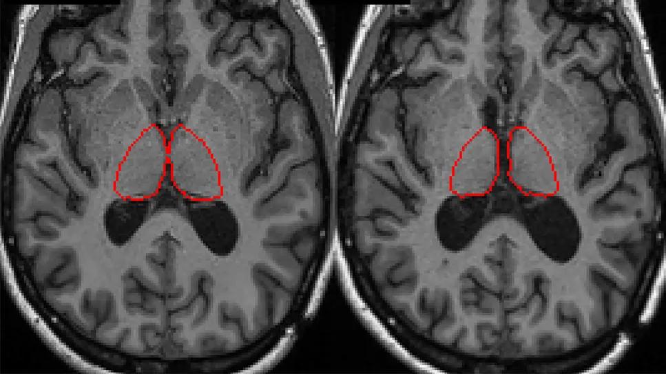
Image content: This image is available to view online.
View image online (https://assets.clevelandclinic.org/transform/228cb07b-63de-4dc4-9991-1f750bb4ba6e/cognitive-profiles-associated-with-thalamic-atrophy-in-ms)
two brain MRIs side by side with red circles on them
Thalamic atrophy is significantly associated with unique neuropsychological testing (NPT) profiles in people with multiple sclerosis (MS), independent of other regional brain quantitative measures. So finds a new study by Cleveland Clinic researchers that leveraged machine learning to link cognitive impairment and neuroimaging biomarkers in MS.
Advertisement
Cleveland Clinic is a non-profit academic medical center. Advertising on our site helps support our mission. We do not endorse non-Cleveland Clinic products or services. Policy
The findings were published in Multiple Sclerosis and Related Disorders (2025;103:10662).
“Using clustering, we identified a profile of neuropsychological testing results in patients with MS that is strongly linked to and possibly driven by thalamic atrophy,” says Moein Amin, MD, a neurologist with Cleveland Clinic’s Mellen Center for Multiple Sclerosis Treatment and Research. “Our results offer novel insights into the application of machine learning to make neuropsychological testing data easier to interpret and incorporate into clinical practice and research.”
Cognitive impairment in MS is common and complex, often reducing quality of life and limiting employment and social functioning. Thalamic volume is known to be correlated with cognition, and thalamic atrophy is associated with neurodegeneration, making thalamic volume a possible biomarker for MS-specific cognitive impairment.
“Thalamic volume and atrophy are well established neuroimaging correlates for disability worsening and cognitive impairment in MS,” Dr. Amin explains. “Regional thalamic atrophy has been linked with specific cognitive domain impairments, including processing speed and memory, in previous studies.”
NPT is useful for evaluating cognitive impairment in patients with MS, but interpretation and integration of all the elements produced from NPT in clinical practice and research is difficult. Prior studies have used clustering to identify homogenous cohorts and previously unrecognized patterns, as well as to define cognitive phenotypes of patients with MS. However, the relationships between these cognitive impairment clusters and phenotypes and longitudinal radiological findings are not well established.
Advertisement
To better explore this area, the Cleveland Clinic researchers undertook a retrospective analysis to evaluate NPT and longitudinal neuroimaging biomarkers in patients with MS using clustering.
Data for the research came from 112 adults with MS who had undergone brain MRI before and after NPT at Cleveland Clinic. Assessments across 19 different domains were collected during NPT. Changes in rates of thalamic atrophy, gray matter atrophy, hippocampal atrophy and T2 lesion volume accumulation were calculated with mixed effects models. These patients’ neuroimaging findings were compared with neuroimaging data from 39 healthy controls who had undergone at least two brain MRI scans; controls did not undergo NPT.

Image content: This image is available to view online.
View image online (https://assets.clevelandclinic.org/transform/688396a4-a5a5-421c-89b5-3d846df81237/cognitive-profiles-associated-with-thalamic-atrophy-in-ms-inset)
Longitudinal brain MRIs demonstrating thalamic atrophy (red-circled regions) over a six-year period. Image courtesy of the Kunio Nakamura lab, Cleveland Clinic.
The participants were divided into training and validation cohorts, and NPT data were condensed, clustered and correlated with longitudinal neuroimaging markers using machine learning. Because of the abundance of variables from the battery of NPT domains and the relatively small sample size, the researchers used principal component (PC) analysis to cluster NPT profiles independent of MRI changes. MRI change rates were subsequently compared across each cluster.
“Machine learning allowed us to reduce the dimensionality of NPT to allow clustering,” says Dr. Amin. “With it, we could more easily interpret the high dimensional data and identify specific cognitive clusters, which can be helpful in personalized decision-making.”
Study participants were predominantly female (71%), and most (80%) had relapsing-remitting MS. Their mean age at NPT was 48 years and their median duration of MS was 12 years.
Advertisement
Compared with healthy controls, study participants with MS had significantly greater rates of change in all MRI measures between their first and subsequent MRIs: thalamic atrophy (P < .001), gray matter atrophy (P < .001), hippocampal atrophy (P = .005) and T2 lesion volume accumulation (P < .001).
Analysis of the training cohort showed that NPT clustering was primarily driven by the first principal component (PC1), to which the most important contributors were measures of processing speed and memory.
The researchers identified two clusters based on PC1, one of which had a significantly greater degree of thalamic atrophy in both the training and validation cohorts and similar rates of change in the other measured quantitative MRI metrics. Significant correlation was found between PC1 and the thalamic atrophy rate, with higher standardized cognitive scores associated with lower rates of atrophy.
“The linear correlation between the first principal component (PC1) and thalamic atrophy rates suggests an important link between thalamic atrophy and selective cognitive impairment,” Dr. Amin says. “The relative magnitude of each contribution is key, as it takes us beyond dichotomized results of individual tests to incorporation of interactions among multiple NPT measures.”
The researchers note that the study’s small sample size did not allow for use of additional clustering methods. As a result, they acknowledge, larger studies with inclusion of longitudinal NPT results are needed to help validate their findings. However, they believe their clustering method may be useful for disentangling various components of cognitive impairment in patients with MS.
Advertisement
“We found a direct correlation between severity of thalamic atrophy — a proposed biomarker of neurodegeneration in MS — and a principal component that had its greatest relative contributions from tests of processing speed and memory,” Dr. Amin concludes. “This result could be particularly important in clinical practice and might ultimately enable more accurate screening, monitoring and evaluation of novel neurotherapeutics for cognitive impairment specific to MS pathology.”
Advertisement
Advertisement

Nearly one-fifth of such cases fulfill 2024 McDonald criteria based on biomarkers

Two research projects aim to enable more personalized MS care in this population

Mixed results from phase 2 CALLIPER trial of novel dual-action compound

A co-author of the new recommendations shares the updates you need to know

Rebound risk is shaped by patient characteristics and mechanism of action of current DMT

First-of-kind prediction model demonstrates high consistency across internal and external validation

Real-world study also finds no significant rise in ocrelizumab-related risk with advanced age

This MRI marker of inflammation can help differentiate MS from mimics early in the disease