Locations:

Clinical complexity demands personalized care strategies
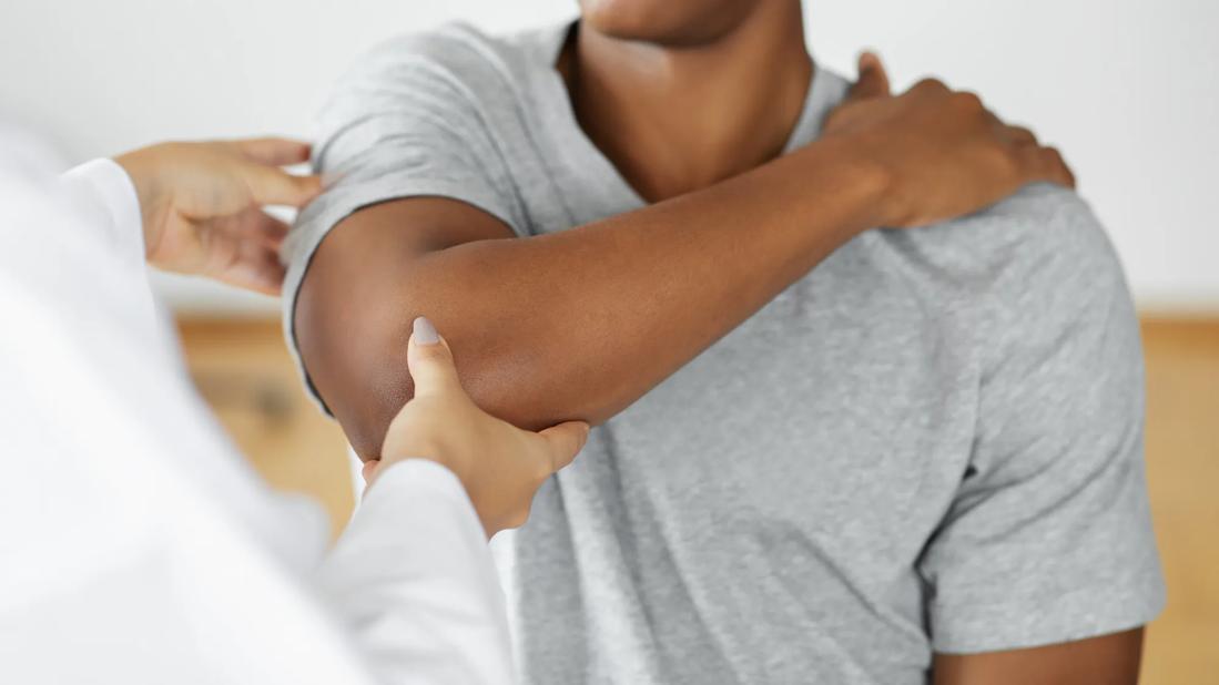
Image content: This image is available to view online.
View image online (https://assets.clevelandclinic.org/transform/a4c74133-8247-4299-88ae-0bfddd8a29f4/elbow-osteochondritis-dissecans-1454566124)
Physician examining patient's elbow
Osteochondritis dissecans (OCD) is a condition in which an area of the bone and cartilage around a joint does not receive a robust enough blood supply to allow microinjuries to heal. Elbow OCD is typically characterized by pain in the lateral part of the elbow, specifically in the capitellum, and the injury is especially common among baseball pitchers and gymnasts after stress from repetitive overhead and loading activities.
Advertisement
Cleveland Clinic is a non-profit academic medical center. Advertising on our site helps support our mission. We do not endorse non-Cleveland Clinic products or services. Policy
"When a young pitcher complains of lateral elbow pain, swelling and loss of motion, I’m always suspicious of elbow OCD," says Paul Saluan, MD, Director for Pediatric and Adolescent Sports Medicine at Cleveland Clinic. “But even if I have my suspicions, elbow OCD is a difficult diagnosis.”
Dr. Saluan says that a focus at Cleveland Clinic is to better understand how to treat elbow OCD more reliably and keep improving outcomes. This has led to research initiatives focusing on the entire injury timeline from presentation to return to play.
“OCD is a condition that we’re seeing more frequently over the past few decades, but it’s difficult to determine whether the incidence rates of OCD have increased or if we’re becoming better at recognizing it,” says Dr. Saluan. “But despite the higher frequency, one medical center won’t see enough OCD cases to be able to have enough data to come away with insights. That’s why Cleveland Clinic has been involved in a multi-institutional collaboration that’s currently focusing on epidemiology, MRI studies and classification.”
Elbow OCD is a combination of several factors that allow a piece of bone or cartilage to become unstable. If untreated, the bone and cartilage can become so unstable that it can become dislodged from its donor site and form a loose fragment.
“This is not a congenital or developmental issue, but it does occur more often in younger athletes due to their unique blood supply to that area of the bone,” says Dr. Saluan.
Patients with elbow OCD often present with lateral elbow pain and may complain of clicking or popping sensations. In more advanced cases, loss of motion and swelling may also be associated.
Advertisement
If elbow OCD is suspected, imaging is typically the primary tool for confirming a diagnosis and treatment recommendations. Typically, a patient will initially have an X-ray followed by MRI.
“X-rays can tell you if there is an OCD there, and an MRI will give you info about the more qualitative aspects of the injury,” says Dr. Saluan. “The MRI can help you determine if it's stable or unstable, if there's cystic change behind it, or if fluid is involved, which would signify instability. But with OCD, like many of these types of injuries, lesion location and size are the two biggest factors involved with clinical decision making.”
Dr. Saluan published research in 2023 that helps provide consensus on some of the most important MRI features to consider when evaluating for capitellar OCD. The group found that lesion containment, the presence or absence of high signal intensity or fluid between the progeny and parent bone, cartilage fissures, lesion size on the sagittal view, and the presence of cysts or cyst-like lesions in the capitellum were the key characteristics.
“The multi-institutional research group that we’re involved with has been examining all of the different factors that could lead to elbow OCD,” says Dr. Saluan. “For instance, we are looking at exposure to smokers since this can constrict small blood vessels and impede the healing process in affected areas. That’s something that one medical center just wouldn’t have enough data on, so the insights we’ve been able to glean from sharing data have been incredibly valuable.”
Advertisement
Once elbow OCD has been confirmed through imaging, there are several factors to consider when making the decision to treat surgically or not.
When assessing a lesion, it is important to consider its size, stability, whether it is chronic or acute, and, most importantly, its location.
“What I’ve found in my research is that with larger lesions that are sitting more laterally, it’s best to reconstruct because the elbow doesn't do well if you only debride,” says Dr. Saluan. “It loses its biomechanical advantage, and the patient ends up with early arthritis.”
“With lesions that are smaller and more centralized, you can just debride those to get the loose fragment out,” he continues. “In those cases, I'm not reconstructing anything. Those patients do well long-term because that lateral pillar of bone, that biomechanical advantage, is maintained, so they don't end up developing significant arthritis.”
Dr. Saluan also notes that younger patients with open growth plates also tend to do better non-surgically since the open growth plates help maintain their range of motion.
For patients who do require surgery, there are a couple of different options. The surgeon may debride the area and either remove or reattach any loose fragments, or they can reconstruct with new cartilage from the patient or a donor.
“If we’re reconstructing an area, one option is to take bone and cartilage from the patient’s knee,” explains Dr. Saluan. “Another option is to get a cadaver graft of cartilage and bone, and that’s typically my preferred method. We’re currently doing a sub-analysis of these two options, and we’re hoping to prove that there’s no significant difference between these two techniques.”
Advertisement
Dr. Saluan also says that with allograft reconstructions, he tends to irrigate the cadaver tissue with pulsatile lavage, which helps clean out the allograft patient’s bone protein. He then soaks the allograft tissue in the recipient patient’s platelet-rich plasma to help speed up the healing process timeline.
“It’s a process where we try to really make the tissue very receptive to taking the implant well,” says Dr. Saluan.
“One of my priorities with patients who have had surgery is to try to get them moving as soon as possible,” says Dr. Saluan. “We want to preserve their range of motion as much as we can. If the elbow doesn’t move for four to six weeks, then they tend to become very stiff, and it’s another battle to get them to move again. The way we do reconstructions is to get the patient stable enough to get them moving from time 0. We just don’t load them, and then by about the fourth or sixth week, we get them involved in physical therapy.”
Dr. Saluan says that oftentimes, the hardest part is that these patients are typically young and active and don’t stop, so getting them to slow down and do activities that don’t stress the elbow can be challenging. But returning to play too early can be horribly detrimental if they're not ready yet. Putting stress through a graft that's not healed yet could cause to fall apart.
However, determining whether a graft is fully healed can be difficult since the healing process is so individualized. Just like during diagnostics, imaging is also typically relied upon to chart a patient’s progress.
Advertisement
“I don’t usually get the first set of X-rays until about three months after a surgery, and then we would do follow-up imaging every two or three months after that,” says Dr. Saluan. “Some patients get repeat MRIs starting at six months to see what kind of interval healing has occurred, but I tend to wait until about a year out. So, there’s variability in determining that, and that’s something our research group is looking at with how to best determine when a graft has healed.”
He notes that while elbow OCD is inherently a complex condition, what makes it much more challenging is the lack of standardization across the injury arc. While diagnostics, imaging and treatments have all improved, the lack of standardization makes injury management inconsistent.
“OCD is a condition that’s extremely individualized,” says Dr. Saluan. “There’s not a ton of available research that can guide appropriate treatment, and that’s something we’ve tried to solve with our multi-institutional research group. We believe our efforts and research can lead to the kinds of diagnostic, treatment, and management recommendations that can push care forward. Managing this condition ultimately depends on patient response, but identifying criteria or signifiers for taking certain approaches to care can improve outcomes and, hopefully, help prevent injuries.”
Advertisement

Protein expression in synovial fluid indicates patients’ immune factors may be involved

Collaboration must cross borders and disciplines

Higher than 5-year rates for prostate cancer and melanoma and about the same as breast cancer

Sinus tracts can occur years later and not near the incision site
Researchers hope it may one day help patients avoid explantation surgery
Symptoms complement one another

Even patients with reported penicillin allergies can receive it without increased complications

Cleveland Clinic’s Global Peak Performance Center and PGA TOUR partnership pair advanced assessment with longitudinal follow-up to enhance clinical decision-making