Locations:

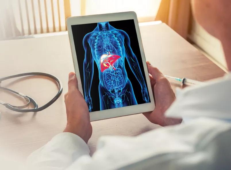
Image content: This image is available to view online.
View image online (https://assets.clevelandclinic.org/transform/e929e12a-aaff-4cea-98d2-43107ece97c1/liver-tumor-article_jpg)
Expanding Treatment Landscape for Gastric Varices
Interventional endoscopists at Cleveland Clinic Weston Hospital have adopted a novel endoscopic ultrasound (EUS)-guided treatment for gastric varices (GV), a common complication in people with liver cirrhosis.
Advertisement
Cleveland Clinic is a non-profit academic medical center. Advertising on our site helps support our mission. We do not endorse non-Cleveland Clinic products or services. Policy
These enlarged veins can occur when scar tissue in the liver blocks blood flow through the portal vein, resulting in portal hypertension. Blood is forced to find new pathways, often through veins in the esophagus and stomach. These usually small veins can become overloaded, causing them to weaken, leak and potentially rupture.
Gastric varices occur in approximately 20% of patients with cirrhosis. They are less common than esophageal varices but are associated with higher mortality (45%).
“Collateral vessels in the stomach tend to be larger and are less prone to bleeding, but when they do bleed, it is much more severe,” explains Tolga Erim, DO, Region Director of Endoscopy and Division Chair of the Department of Gastroenterology, Hepatology and Nutrition for Cleveland Clinic in Florida. “Fortunately, we have an increasingly wide array of treatment options to offer patients with high-risk bleeds.”
Unlike esophageal varices, gastric varices are too large to be managed with endoscopic variceal ligation, a banding procedure used to occlude the vessels.
Endoscopic cyanoacrylate glue injection (ECI) was introduced in the 1980s and has become the most widely used method to definitively treat GV. This technique has been shown to prevent early and late rebleeding, but it is not without risk. Embolic incidents are clinically evident in about 1% of cases.
“Systemic glue embolization leading to pulmonary embolus or stroke is a rare occurrence but it can have devasting consequences,” says Dr. Erim.
Advertisement
More recently, the addition of endoscopic ultrasound guidance has improved the precision and safety of variceal treatment. Dr. Erim notes the technique allows for better placement of the fine needle aspiration needle with the added benefit of real-time Doppler flow assessment to confirm hemostasis.
Today a number of EUS–guided options are used to treat GV, including glue injection, coil embolization, and coil embolization combined with glue injection. These EUS-guided approaches can achieve high obliteration rates with minimum reintervention and fewer adverse events, according to the literature.
One single-center, parallel-randomized controlled trial also found that “EUS-guided coil embolization with cyanoacrylate injection achieved excellent clinical success with lower rates of rebleeding and reintervention than coil treatment alone.”
“An advantage of the coil plus glue approach is the reduced risk of systemic embolization,” reports Dr. Erim. “Not only do the coils reduce blood flow, they also act as scaffolding for the glue to keep it in place.”
A newer method combines EUS-guided coil embolization with hemostatic absorbable gelatin sponge (AGS) injection in place of cyanoacrylate. This technique was recently used by Dr. Erim and his team at Weston Hospital. “By using gelatin sponge instead of glue we hope to further reduce the risk of adverse events as seen in early research,” Dr. Erim adds.
In addition to the many endoscopic treatments for GV, there are also endovascular management options performed by interventional radiologists. Transjugular intrahepatic portosystemic shunt (TIPS) and balloon-occluded retrograde transvenous obliteration (BRTO) are used electively or emergently and have comparable clinical outcomes, according to practice guidelines published in 2021.
Advertisement
TIPS is a percutaneous procedure that creates a shunt between the portal vein and hepatic vein within the liver. It’s designed to reduce portal pressure and redirect blood flow away from esophageal and gastric varices.
During a BRTO procedure, a balloon catheter is inserted into a draining vein of the GV where it is inflated to block blood flow. A sclerosant is then injected into the varix to cause vessel sclerosis. Unlike TIPS, this approach causes hemostasis while increasing portal pressure.
“There are many factors that can determine which treatment option or combination of treatments is most appropriate,” says Dr. Erim, noting practice standards are limited. “That’s why AGA guidelines recommend a multidisciplinary approach for managing gastroesophageal varices.”
Earlier this year, Dr. Erim performed Weston Hospital’s first EUS-guided coil embolization combined with AGS injection to stop acute variceal bleeding in a 77-year-old female diagnosed with cirrhotic portal hypertension.
Prior to transfer to the hospital, the patient underwent a TIPS placement that failed to achieve primary hemostasis. “In this case, the patient was too unstable for BRTO, and the bleeding was too extensive for a cyanoacrylate injection alone,” says Dr. Erim.
Instead, Dr. Erim used a transgastric approach to place five embolization coils into the varix, followed by injection of gelatin sponge mixed with saline. There were no immediate complications and the patient required only one unit of blood during the procedure. Primary hemostasis was achieved, and complete obliteration of the GV was observed during the procedure.
Advertisement
“The patient did well, and we were very pleased with the outcomes in this case,” shares Dr. Erim. “We plan to use this approach more often in the future.”
As a tertiary care center, Weston Hospital has a team of gastroenterologists, endoscopists, interventional radiologists and surgeons who are able to offer multiple therapeutic options for gastric varices. The hospital is also home to a liver transplant program, providing the only cure for end-stage liver disease.
More than 630,000 adults in the United States, or about 1 in 400, are estimated to have cirrhosis. Some of the most common causes are viral hepatitis, alcohol use, and nonalcoholic fatty liver disease (NAFLD).
According to the American Liver Foundation, about 24% of U.S. adults are estimated to have NAFLD and a quarter of them will progress to nonalcoholic steatohepatitis, placing them at greater risk of developing cirrhosis.
“The role of obesity and type 2 diabetes in the development of fatty liver disease is contributing to an increase in cirrhosis, especially here in the United States,” notes Dr. Erim. “As a result, we are likely to see more patients with gastric varices.”
Advertisement
Advertisement

Early recognition and intervention recommended in cubital tunnel syndrome

Manpreet (Meena) Bedi, MD, named Division Chair of Radiation Oncology

AR-assisted navigation is closing the gap between surgical planning and implant placement

Evidence shows early evaluation improves survival and quality of life – yet many eligible patients are referred too late

2026 ADA Standards of Care promote holistic, multisystem management
Cleveland Clinic nephrologist in Florida addresses changes in clinical practice

Surveillance platform supports community clinicians and public health monitoring

Benefits include improved clinical outcomes and lower healthcare costs