Locations:

Sports medicine pioneer John Bergfeld, MD, shares how orthopaedics has changed since doing his first ACL repair in 1970
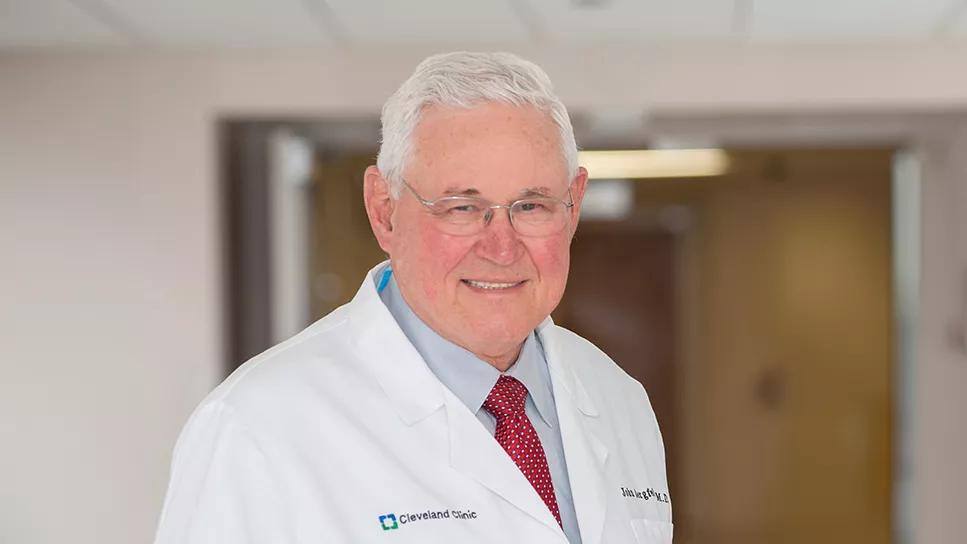
Image content: This image is available to view online.
View image online (https://assets.clevelandclinic.org/transform/306b898d-cd93-4ab8-807b-cc0df695c451/john-bergfeld)
John Bergfeld, MD
Long before John A. Bergfeld, MD, became a leader of sports medicine at Cleveland Clinic and a pillar of the orthopaedic profession, he was Chief of Orthopaedics at the U.S. Naval Hospital in Annapolis, Maryland, and team physician for the U.S. Naval Academy.
Advertisement
Cleveland Clinic is a non-profit academic medical center. Advertising on our site helps support our mission. We do not endorse non-Cleveland Clinic products or services. Policy
He saw numerous midshipmen with the same mysterious knee injury. Often, they’d land after a jump, “feel a snap” and then report disabling knee pain. Within 24 hours, they’d develop significant effusion.
“I didn’t know what it was, but I thought the Army had to be seeing the same thing,” Dr. Bergfeld says. “So, before the Army-Navy football game in 1970, I went over to the Army sideline and asked to talk to their doctor. He refused to talk to anybody from Navy before the game! When I returned to Annapolis after the game, I had my corpsman call West Point every day until I finally got a call back from their team doctor, John Feagin. He’s the one who told me that what I had been seeing were torn cruciate ligaments.”
Dr. Feagin, who later became renowned as an orthopaedic sports medicine pioneer, explained how the ligaments were being repaired at West Point. That inspired Dr. Bergfeld to try it too, by opening the knee and suturing the torn ligament.

Image content: This image is available to view online.
View image online (https://assets.clevelandclinic.org/transform/9c5ba862-ec60-4558-9c84-c8c2106bbe27/Bergfeld-examines-athlete-torn-ACL)
Dr. Bergfeld (in white uniform at right) examines a Navy football player with a knee injury. (Photo taken in 1970.)
Orthopaedic leaders at the time didn’t look favorably on cruciate ligament repair. It was an extensive procedure, and patients were advised not to return to playing sports. A 1972 article in The Medical Post claimed the surgery was “of little help for cruciate ligament injury.”
At the 1972 American Academy of Orthopaedic Surgeons meeting, Dr. Feagin presented the outcomes of West Point cadets two years after primary cruciate ligament repair. More than 80% of them were doing well. Proud that he had been doing the same surgeries in Annapolis, Dr. Bergfeld turned excitedly to tell a seasoned colleague, but shrank back when the colleague began lambasting the presentation.
Advertisement
Three years later, Dr. Feagin’s outcomes didn’t look as good. At five-year follow-up, 38% of the West Point cadets had impaired normal activity, 53% had significant reinjuries, 70% had pain and swelling, and 94% had knee instability.
“In other words, the ligament repair hadn’t worked,” Dr. Bergfeld says. “It confirmed the saying ‘nothing like good follow-up to ruin good results.’ My first lesson in orthopaedics was: Don’t adopt new treatments right away unless you’re the one researching them. Two-year results are unreliable.”
Why had some of the operations initially been successful? Likely because the patients hadn’t yet regained full range of motion, says Dr. Bergfeld. After the operations, patients wore a long leg cast for three weeks, leading to stress deprivation and muscle atrophy. As long as the patients never fully extended their repaired knee, maintaining a five-degree flexion contracture, the ligament repair seemed sufficient.
Dr. Bergfeld admits that he was a victim of the orthopaedic sports medicine triad, which he defines as a:
Advertisement

Image content: This image is available to view online.
View image online (https://assets.clevelandclinic.org/transform/21f63be5-764b-4691-bc90-5837c481c285/pes-anserinus-transplantation)
Dr. Bergfeld demonstrates pes anserinus transplantation, an early surgical treatment for knee instability, which involved rotating the combined tendons 180 degrees. (Photo taken in 1972.)
In 1972, a few years after the introduction of surgical treatment for AMRI, the pivot shift test was described, leading to the diagnosis of anterior lateral rotary instability (ALRI). This developed the understanding that unstable knees had both anterior medial and anterior lateral rotary instability — thus requiring two surgical procedures, one on each side.

Image content: This image is available to view online.
View image online (https://assets.clevelandclinic.org/transform/71a0ab48-fa16-4064-9e08-8ecd0ee1f577/medial-and-lateral-transfer-surgeries)
Dr. Bergfeld demonstrates medial transfer and lateral transfer, two procedures required to address instability in one knee. (Photo taken in 1975.)
“They were gigantic incisions. It took as much time to close them as it did for the repair,” Dr. Bergfeld says. “We knew that the anterior cruciate ligament [ACL] was torn as well, but we didn’t pay attention to it. The dominant voices in orthopaedics at the time said that an ACL didn’t need to be repaired as long as we repaired the meniscus and capsular pathology, so that’s what we believed.”
That changed around 1976, when Ejnar Eriksson, MD, from Sweden and Kurt Franke, MD, from East Germany simultaneously published their clinical experiences with cruciate ligament reconstruction using the patellar tendon. Dr. Bergfeld was invited behind the Iron Curtain to hear a talk by Dr. Franke, who stressed the importance of reproducing normal anatomy.
“Years earlier, U.S. surgeon Kenneth Jones had published on using the patellar tendon to repair the cruciate ligament, but he reported 60% poor results,” Dr. Bergfeld says. “As a result, nobody in the U.S. paid attention to reconstruction with the patellar tendon. It turned out that he was inserting the tendon in the wrong place. Had he done it to be anatomically correct, we’d likely have been doing cruciate ligament reconstruction with the patellar tendon 15 years sooner.”
Advertisement
Regardless, thanks to the trailblazing work by Dr. Jones and modifications by Drs. Eriksson and Franke, U.S. orthopaedic surgeons began performing cruciate ligament reconstruction as well — but only in addition to the medial and lateral procedures.
“These surgeries required multiple large incisions and took hours,” Dr. Bergfeld says. “However, once we started doing the ACL reconstructions, we started wondering about the role of the extra-articular procedures. Maybe we didn’t need to be doing these great big operations if we just repaired the ACL.”
So, in 1989, Dr. Bergfeld, then Chair of the Foundation for Sports Medicine Education and Research, gathered 28 prominent U.S. orthopaedic surgeons to discuss the topic. Three days later, they conceded that nothing in the literature supported performing extra-articular procedures on patients with torn ACLs.
“We had been doing them every day for 10 years, but finally realized that there was no evidence that intra-articular ACL substitution was improved by adding an extra-articular procedure,” Dr. Bergfeld says. “Out of the 28 surgeons, 24 of us began doing intra-articular procedures alone — primary ACL reconstructions.”
As primary ACL reconstruction became prominent, surgeons began exploring alternatives to using the patellar tendon, to avoid harvesting grafts. Carbon fiber was one option, but that stopped when particles began showing up in patients’ inguinal lymph nodes, notes Dr. Bergfeld. Allografts were another option, until patients started developing synovitis, caused by the ethylene oxide used to sterilize the grafts. Bovine tendon xenografts and artificial ACL prostheses failed as well.
Advertisement
“Since then, we’ve still been unable to develop a successful artificial ACL,” Dr. Bergfeld says. “Ejnar Eriksson was right when he said ‘Artificial ACL substitutes are like shoestrings. Eventually they all break.’”
Surgical failures like these are more often avoided today, he adds, thanks to oversight by the U.S. Food and Drug Administration and institutional review boards.
The advent of arthroscopy led to much smaller incisions in ACL reconstruction. No longer were big incisions needed to harvest grafts.

Image content: This image is available to view online.
View image online (https://assets.clevelandclinic.org/transform/de567253-c6fd-4a18-bb73-42d490548a99/patellar-tendon-harvesting-for-ACL-reconstruction)
Dr. Bergfeld demonstrates the difference in incision size when harvesting a patellar tendon graft with conventional surgery (left) versus arthroscopic surgery (right).
Next came single-incision ACL reconstruction, a technically difficult procedure for graft placement.
“When we went from double- to single-incision surgery, many surgeons reverted to putting grafts in the wrong spot on the femur — the same problem we saw in early surgeries,” Dr. Bergfeld says.
At that time, most of Dr. Bergfeld’s surgeries were ACL revisions. In 1998, he and his counterparts at the University of Pittsburgh and the Hospital for Special Surgery found that up to 60% of their revision surgeries were due to technical errors, such as improper graft placement.
Until Dr. Bergfeld retired in 2024, he continued to perform ACL repair with two incisions to ensure proper graft placement.
“Single-incision surgery was becoming popular, but few surgeons at the time could do it correctly,” he says. “They showed the technique at an academy meeting, and some went home and just did it without really knowing how.”
When double-bundle reconstruction (using anteromedial and posterolateral bundles) was introduced, it was a similar situation. The technically challenging procedure required two drill holes in the area of the femur where it was difficult to place even one, explains Dr. Bergfeld.
“Freddie Fu, the champion of double-bundle reconstruction, said even he couldn’t do the surgery in many cases. The consensus of multiple studies is that there’s little to no difference in clinical outcome between double- and single-bundle surgeries. One drill hole placed properly works just as well and is much easier to do.”
The takeaway for surgeons is ensuring they know the proper way to perform a procedure before offering it to patients.
“Maybe visit the surgeon who’s developing it,” Dr. Bergfeld advises. “And always pay attention to what team doctors are doing. Team doctors get 100% follow-up from their athletes in the training room, so you can trust the procedures they are doing.”
Advanced technology has greatly improved outcomes for patients with torn ACLs.
“Surgeons today are so well trained technically,” Dr. Bergfeld says. “And they can instantly access literature and other resources. Technology is a giant asset to both doctors and patients. But don’t let that technology take the place of caring for patients.
“Some surgeons complete the surgery and turn the patient over to another provider for follow-up. You don’t want your patients to say, ‘I went to this surgeon for my operation and then never saw them again.’ Remember why you wanted to be a doctor instead of a technician. Take care of your patients.
“As one old doctor once said, ‘Just walk in, put your hand on the patient’s knee and look them in the eye. It’ll pay you big dividends.’”
Advertisement

Largest cohorts to date reveal low rates of major complications

Clot substitute helps rejoin the stumps of a torn ligament

Reducing prescriptions may help keep unused medication out of the community

Multidisciplinary care can make arthroplasty a safe option even for patients with low ejection fraction

Self-care may be just as effective for some patients

Most return to the same sport at the same level of intensity

Higher than 5-year rates for prostate cancer and melanoma and about the same as breast cancer

Insights to help orthopaedic practices comply with the 2025 CMS mandate