Locations:





How careful measurement, provocative testing and repeat assessment can maximize the diagnostic value of PAC
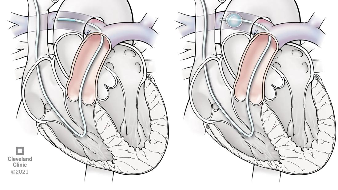
Image content: This image is available to view online.
View image online (https://assets.clevelandclinic.org/transform/8119b072-7b4b-4875-a218-8a72bbd6dcc9/pulmonary-artery-catheter)
Pulmonary artery catheterization illustration
Written by Matthew T. Siuba, DO, MS and Adriano R. Tonelli, MD, MSc
Advertisement
Cleveland Clinic is a non-profit academic medical center. Advertising on our site helps support our mission. We do not endorse non-Cleveland Clinic products or services. Policy
Pulmonary artery catheterization (PAC), sometimes called Swan-Ganz catheterization in honor of its creators, is the most widely used invasive method to assess patients with cardiopulmonary disorders in both outpatient and inpatient settings, including the intensive care unit (ICU). Because the catheter is advanced through the right ventricle into the pulmonary artery where it sits, the term “PAC” more specifically describes its position than the more commonly used term “right heart catheterization.”
Over twenty years ago, two large randomized clinical trials (PAC-Man trial and ESCAPE trial) in ICU patient populations showed no improvement in outcomes with the use of PAC compared to usual care. These results led many clinicians to shy away from PAC, ultimately leading to a decrease in catheter placement skills as well as reduced ability to interpret the hemodynamic data it provides.
However, PAC remains a cornerstone for assessing patients with suspected or confirmed pulmonary hypertension (PH). Additionally, more recent observational cohorts have shown an association with improved outcomes in patients with cardiogenic shock when comprehensive PAC assessment is used. This has led to new clinical trials investigating this question. Intensive care societies also recommend PAC in patients with complex circulatory shock states, especially when respiratory failure is also present.
Expert use of PAC requires that all invasive data is carefully and consistently acquired. Because the catheter is fluid-filled, proper setup before placement is essential: pressure transducers must be zeroed and leveled at the right atrium, which is typically approximated by the mid-axillary line at the fourth intercostal space. Transducers should be assessed for over- and under-damping, which can affect the accurate measurement of systolic and diastolic pressures. To minimize the effect of intrathoracic pressure changes, all pressures should be measured at the end of expiration, when intrathoracic pressure is generally negligible, regardless of whether the patient is breathing spontaneously or mechanically ventilated.
Advertisement
For a comprehensive hemodynamic evaluation, all available pressures should be measured, including the right atrial, right ventricular pressure, pulmonary artery and pulmonary artery wedge pressures (PAWP). Experienced operators learn the characteristic waveform morphologies to recognize the differences and extract the most information. Although waveform characteristics provide information about atrial and ventricular function, a more detailed discussion is beyond the scope of this article.
Accurate PAWP measurement is essential to estimate left atrial pressure properly. This measurement influences patient management in multiple ways. In addition to providing a direct assessment of filling pressure, it is also used to calculate metrics such as pulmonary vascular resistance (PVR), which can affect PH hemodynamic classification. A PAWP is more likely to be accurate when all the following criteria are met:
Although pulmonary vascular pressures are measured at end-expiration, certain conditions — including obesity and air trapping from obstructive lung disease — can result in a positive intrathoracic pressure during this part of the respiratory cycle. In these cases, esophageal manometry can estimate intrathoracic pressure, which can then be used to adjust the pulmonary vascular pressures. When this is not available, some societies recommend using mean (machine-averaged across the respiratory cycle) values for pressures rather than end-expiratory determinations.
Advertisement
It is critical to assess the measured pressures from the PAC in context with accurate cardiac output (CO) measurement. CO is also necessary for calculating derived values like PVR or pulmonary artery compliance. Indirect or estimated Fick and thermodilution are the two most common methods for CO estimation. However, both methods are prone to imprecision and should be interpreted cautiously and in clinical context.
Overall, most guidelines recommend thermodilution instead of indirect Fick CO. Thermodilution is less prone to calculation errors than the gold-standard direct, or measured, Fick CO method, which requires measurement of oxygen consumption with a metabolic cart that is not available in most catheterization laboratories. When thermodilution CO determination is used, measurements should be performed in triplicate with < 10% variation to minimize variability. Notably, the presence of tricuspid regurgitation has not been convincingly shown to alter the accuracy of CO measured by thermodilution. Direct Fick CO should be considered when precision and accuracy of CO are essential, such as those near cut-off points for cardiac index (e.g. 2.2 and 4 L/min/m2).
Sometimes the patient’s resting state doesn’t accurately represent their disease state under normal life situations. This is especially true in patients with dyspnea on exertion, or well-compensated patients with underlying heart failure and pulmonary hypertension. In these cases, dynamic testing during PAC can improve diagnostic accuracy.
Advertisement
Inhaled pulmonary vasodilators such as nitric oxide are often used to assess vasoreactivity of the pulmonary arterial circulation in patients with precapillary PH. In patients with a high pre-test probability of heart failure with preserved ejection fraction (HFpEF) but normal PAWP, a fluid challenge can help detect occult HFpEF, since the PAWP typically increases above 18 mmHg.
Inotropic agents such as dobutamine can be used to assess ventricular contractile reserve. This may be useful for risk stratification of patients with heart failure or PH. This hemodynamic provocation has also been used to risk stratify patients for high-risk surgeries such as liver transplantation.
Many patients experience cardiopulmonary symptoms during exertion, so exercise testing during PAC can help reproduce those symptoms and identify the underlying mechanism. Exercise PAC can be done with or without cardiopulmonary exercise testing, which together is called invasive CPET (iCPET). This can help identify patients with exercise-PH, exercise-HFpEF, and preload insufficiency, among other conditions.
Remember that PAC is a diagnostic tool that should be used for re-assessment too! Whether the patient is receiving treatment instantly in the ICU, or longitudinally in the outpatient setting, repeat assessment with PAC is essential to assess treatment response. If a patient is receiving vasoactive medications or fluids in the ICU, repeat PAC determinations after treatment to guide subsequent management. If an outpatient is started on medications for heart failure or PH, repeat assessment at clinician-determined intervals can assess response and adjust treatment when necessary.
Advertisement
To maximize the diagnostic value of PAC in our laboratory, we offer the following assessments:
Advertisement

The benefits of multiarterial grafting are possible without opening the chest
CMR-CLIP outperforms general AI tools; may one day expand patient access to CMR

An infographic-style overview of our volumes and outcomes

A closer look at the impact on procedures and patient outcomes

Medical and surgical perspectives on current and emerging uses of ECMO and Impella
A scannable recap of our latest data in these clinical areas
Least-invasive open-heart AVR option to date yielded rapid recovery in all cases
How two Cleveland Clinic alliance hospitals systematically elevated echo standards
 Search