Locations:

A closer look at the impact on procedures and patient outcomes
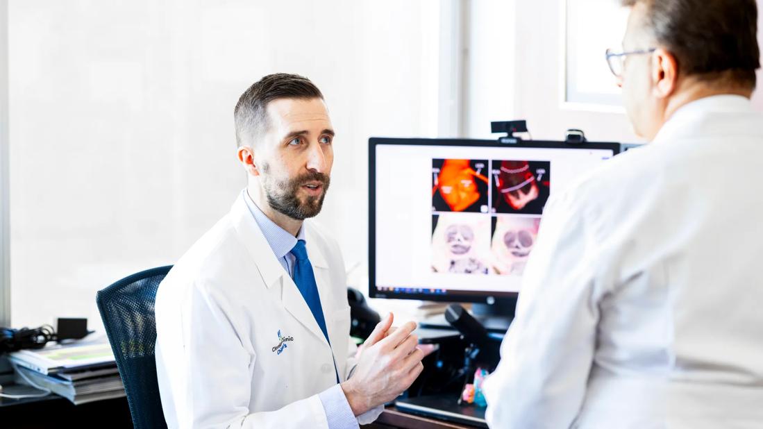
Image content: This image is available to view online.
View image online (https://assets.clevelandclinic.org/transform/d150884a-d316-4c58-a757-862b603b2e31/CHP_5526166_01-27-25_028_LDJ)
Dr. Tretter in conversation with Dr. Najm
Recent developments in cardiac imaging, combined with an enhanced understanding of cardiac anatomy in relation to the conduction system and surrounding structures, are providing a more comprehensive framework for interventional cardiologists and cardiac surgeons.
Advertisement
Cleveland Clinic is a non-profit academic medical center. Advertising on our site helps support our mission. We do not endorse non-Cleveland Clinic products or services. Policy
The stakes are high: damage to the conduction system can occur in upwards of 15% of aortic valve or left ventricular outflow tract surgeries and results in the need for a permanent pacemaker following surgery.
Justin Tretter, MD, a pediatric cardiologist, advanced imager, and one of the few cardiac anatomists in the world, has been at the helm of this work. He’s been leading histological and anatomic investigations into the conduction system and adjacent cardiac structures for more than a decade.
“My work has always been guided by a bench-to-bedside approach, specifically, through the translation of anatomical and developmental findings towards guiding cardiac imaging evaluation to improve clinical care for patients with cardiac anomalies,” he says.
Early in his career, Dr. Tretter trained with renowned cardiac morphologist Robert H. Anderson, MD, PhD (Hon), in London. He utilized developmental and anatomical research methods to understand normal and abnormal cardiac anatomy in autopsied heart specimens. These investigations gave way to discoveries in aortic valve anatomy that would shape his clinical practice and research program.
In healthy hearts, he observed normal variation in the rotational position of the aortic root that correlated with the underlying central fibrous body, a group of structures related to the conduction system.
“That raised a lot of questions for me, as an anatomist, cardiac imager and cardiologist,” he notes. “I wondered how variation in the normal heart affects the valve function, physiology and hemodynamics, not to mention the adjacent conduction system,” he says. “Furthermore, if this variation is significant,” he surmised, “it may have implications for any type of surgery or intervention on the aortic valve or its surrounding structures.”
Advertisement
Dr. Tretter calls the historical literature on the conduction system “important and valid” but missing a key component: a 3D understanding of its position relative to the aortic root and other cardiac structures. The 2D histological piece of the puzzle was limited but critical, as it was the only way to visualize the components of the conduction system and translate them back into the 3D heart.
He again collaborated with his mentor, Dr. Anderson, in London, as well as with Dr. Damián Sánchez-Quintana, an anatomist in Spain, and other experts from around the world.
“This provided a framework for predicting the location of the atrioventricular conduction axis relative to the aortic root by clinical imaging,” says Dr. Tretter. Through this work, which has been validated and reproduced, experts have concluded that its anatomical location is both highly variable and also predictable when its association with surrounding structures is correctly interpreted.
Over the last several years, the advent of hierarchical phase-contrast tomography (HiP-CT), a technology that provides a cellular-scale view of intact human organs with the ability for 3D reconstructions, has been a game-changer. The imaging tool upends the constraints imposed by a 2D visualization of the conduction system and is paving the way for a new era in understanding of cardiac structures.
Using open-access scanned autopsied heart specimen datasets made available by the European Synchrotron Radiation Facility, Dr. Tretter and colleagues analyzed these specimens with the HiP-CT technology and described their findings in Heart Rhythm.
Advertisement
“It scans to the 20 micron-level spatial resolution, and so, for the first time, we now have a 3D dataset of a heart and can visualize all the structures and variation in the conduction system tissue,” he says, likening it to histology but in 3D.
Dr. Tretter and his colleagues then segmented the first accurate 3D reconstruction of a human heart atrioventricular conduction axis from this same dataset. They published this report in Arrhythmia & Electrophysiology Review. This understanding supported Dr. Tretter and his collaborators in creating a standardized method to estimate the 3D anatomy of the conduction system relative to anatomical landmarks in patients using clinical cardiac CT. These methods were published in Heart Rhythm. Dr. Tretter also trained bioengineers to make the same assessment, and they blinded the study, showing that this standardized method is highly reproducible.
“We've established a basis for how a clinical cardiologist or imager can estimate the course of the atrioventricular conduction axis,” he explains.

Image content: This image is available to view online.
View image online (https://assets.clevelandclinic.org/transform/37e373a6-482e-43e2-8f4b-92b4bf23e165/conduction-system)
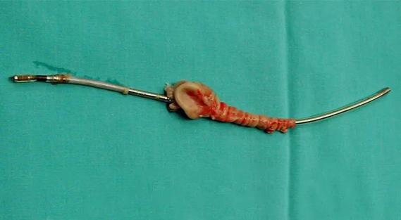
Figure. 3D computed tomography reconstruction of the left ventricle and aortic valve with the atrioventricular conduction axis superimposed. This reconstruction demonstrates estimation of the atrioventricular conduction axis from atrioventricular node (Point A) to His bundle course (Point B) to left bundle branch origin (Point C). This estimated course is then used by the cardiac surgeon to avoid its damage during any aortic valve and left ventricular outflow tract surgery.
These studies have provided a framework for interventional cardiology teams performing transcatheter aortic valve replacement (TAVR), transcatheter mitral valve replacement (TMVR) and transcatheter tricuspid valve replacement (TTVR) to avoid the conduction system as well as for electrophysiologists performing cardiac pacing procedures that target the conduction system.
Dr. Tretter has partnered with CARA Medical to create innovative software solutions that noninvasively simulate the cardiac conduction system to augment fluoroscopy during adult acquired structural heart and cardiac pacing interventions. He is helping to lead clinical trials to evaluate this technology on a global scale. This technology has received FDA breakthrough designation and is currently being trialed at multiple leading centers in North America, Europe, Asia and Australia.
Advertisement
Through this partnership, the team implemented the technology to evaluate pacemaker placement, demonstrating that when the pacing lead is placed within the His bundle or left bundle branch trunk, as opposed to outside of the conduction system within the ventricular septum muscle, patients had improved outcomes. They published these findings in Heart Rhythm and were selected to present the late-breaking clinical trial at the Heart Rhythm Society meeting in May 2025.
More recently, working with an adult cardiac interventional team in Freiburg, Germany, they demonstrated that computed tomography angiography (CTA)-based conduction system simulation may offer an improved patient-specific TAVR deployment strategy, avoiding conduction system damage and the need for a pacemaker. They published these findings in Heart Rhythm. Multiple ongoing studies are now evaluating the use of this approach for real-time augmented fluoroscopic guidance in both structural heart interventions and cardiac pacing procedures.
The team within Cleveland Clinic Children’s Congenital Valve Procedural Planning Program, where Dr. Tretter serves as co-director alongside cardiothoracic surgeon Hani Najm, MD, has adopted similar anatomical and advanced imaging approaches. These techniques guide procedural planning efforts and support better patient outcomes in those undergoing congenital valve surgeries.
“Over the past three years, we’ve performed detailed CT assessments for over 100 patients undergoing congenital aortic valve surgery,” he says. “While our primary focus in obtaining these CTs is to create a personalized blueprint for surgical repair or replacement, this has also allowed us to estimate the location of the conduction system and avoid heart block, reducing the rate from the national average of approximately 10%, down to near zero percent in our patients.”
Advertisement
The team plans to publish their center’s three-year experience soon. Dr. Tretter adds that he hopes it will raise the bar for the standard of care in congenital valve surgeries. They also hope to expand its use for those outside of the congenital heart valve disease population to include patients with other forms of complex congenital heart disease at high risk for conduction system damage.
Advertisement

CMR-CLIP outperforms general AI tools; may one day expand patient access to CMR

Medical and surgical perspectives on current and emerging uses of ECMO and Impella

Least-invasive open-heart AVR option to date yielded rapid recovery in all cases

How two Cleveland Clinic alliance hospitals systematically elevated echo standards

As use for atrial arrhythmias surges, studies turn to ventricular arrhythmias

Launch of the tool promises to reshape quality assessment across the specialty
Lead dwell time and manufacturer emerge as independent predictors of success in registry study

The case for a thoughtful approach to CTO and minimally invasive options for CABG