Locations:

Benefits include reduced pain, earlier mobilization and more likely discharge to home
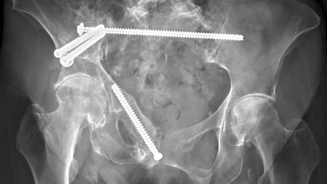
Image content: This image is available to view online.
View image online (https://assets.clevelandclinic.org/transform/675231fa-0eec-4879-ac9b-3504d86ebb50/24-ORI-5289458-CQD-inset4)
X-ray of pelvic ring fixation
By Colin M. Baker, DO; Jared A. Warren, DO; Nathan W. Mesko, MD; and Cesar Cereijo, DO
Advertisement
Cleveland Clinic is a non-profit academic medical center. Advertising on our site helps support our mission. We do not endorse non-Cleveland Clinic products or services. Policy
Pelvic ring fractures are common within our active, aging population. Recently, pathologic pelvic ring and acetabular fractures have been gaining attention in the cancer population, where metastatic disease can disrupt pelvic ring architecture.1,2
Pelvic ring injuries and disruptions are associated with significant pain that worsens with ambulation and can lead to detrimental long-term outcomes. Patients who have these injuries are at risk of prolonged immobilization, which increases the likelihood of requiring ambulatory aids, reduces independence and increases morbidity.3
In the trauma population, it is generally accepted that unstable fractures and fractures with significant displacement are indicated for surgical intervention. Lower-energy, mechanically stable fractures often are treated nonoperatively. In the metastatic disease population, metastatic disease in the pelvis often is treated with radiation alone.
However, with new surgical techniques involving pre-emptive fixation of an impending fracture, we can provide quicker and less difficult recoveries, dramatically improving patients’ quality of life. Data supporting percutaneous pelvic ring fixation surgery are growing. Research indicates reduced pain, earlier mobilization, higher chance of discharge to home and lower risk of mortality at one year following pelvic ring fixation surgery.4
The use of surgical fixation in pelvic ring fractures in older adults remains a debated topic within the orthopaedic community, but as surgeons become more familiar with this minimally invasive technique, more patients will benefit from easier recoveries.
Advertisement
Percutaneous surgical fixation in pelvic ring injuries has been implemented by some surgeons in the trauma and musculoskeletal oncology fields, although its use is not widespread.
This intervention involves small incisions at the lateral and posterior aspect of the hips followed by placement of guidewires for the eventual insertion of large cannulated screws. Photodynamic balloons help navigate bone corridors that require less rigid fixation options.
The number and location of screws depends on fracture pattern and patient symptoms. Posterior pelvic ring fixation using iliosacral and transsacral screws, through the articulation between the sacrum and the ilium, recreates the stability provided by the robust sacroiliac ligaments and is prioritized in this procedure.
Percutaneous pelvic ring fixation is often beneficial in older patients who sustain fragility fractures, such as pelvic fractures after a ground-level fall. Conventional open surgical fixation in older adults with frailty is associated with significant morbidity and mortality. As such, percutaneous fixation can be a welcome alternative.

Image content: This image is available to view online.
View image online (https://assets.clevelandclinic.org/transform/5aca5430-6457-44b2-9e10-50d093961e4f/24-ORI-5289458-CQD-inset1)
AP pelvis radiograph demonstrating right-sided transiliac fracture with ipsilateral superior and inferior pubic rami fractures in a patient after a ground-level fall.

Image content: This image is available to view online.
View image online (https://assets.clevelandclinic.org/transform/38f71505-9f07-4a0b-b2d1-6913f2d538a5/24-ORI-5289458-CQD-inset2)
Axial CT scan demonstrating displaced right-sided iliac wing fracture.

Image content: This image is available to view online.
View image online (https://assets.clevelandclinic.org/transform/21856655-3b72-420d-aa1a-db00760f5903/24-ORI-5289458-CQD-inset3)
3D reconstruction CT scan demonstrating right-sided iliac wing fracture and ipsilateral pubic rami fractures.
For example, after reduction and fixation of an iliac wing fracture, we can perform posterior pelvic ring fixation with a transsacral screw anchored across the S1 sacral body and a retrograde superior rami screw placed for stabilization of the anterior column.
Initially after surgery, patients’ weight-bearing may need to be protected, depending on extent of the injuries. Rehabilitation in a nursing facility and/or with in-home therapy is often recommended. It is common for patients to progress to weight-bearing as tolerated and eventually return to baseline, including ambulating and living independently at home.
Advertisement

Image content: This image is available to view online.
View image online (https://assets.clevelandclinic.org/transform/675231fa-0eec-4879-ac9b-3504d86ebb50/24-ORI-5289458-CQD-inset4)
AP pelvis radiograph of a patient eight months after pelvic fixation. Hardware appears stable with previous pelvic ring fractures healed.
Fragility fractures may be the most common reason for providing patients with percutaneous pelvic ring fixation, but it’s not the only one. Below are two cases highlighting additional scenarios in which percutaneous pelvic ring fixation has been used with good results.
Approximately six years before presenting to the orthopaedic service at Cleveland Clinic, a patient older than age 70 underwent approximately 30 rounds of radiation therapy as part of oncologic treatment. The patient developed worsening atraumatic anterior and posterior pelvic and bilateral hip pain, causing them to rely on a walker for ambulation.
The patient was found to have significant radiation osteitis with associated insufficiency fractures of both the left and right hemipelvis involving the anterior and posterior pelvic ring.

Image content: This image is available to view online.
View image online (https://assets.clevelandclinic.org/transform/4c98c5b7-8af1-4e96-9264-656be946d875/24-ORI-5289458-CQD-inset5)
AP pelvic radiograph demonstrating diffuse sclerotic changes within the pelvis secondary to radiation. There also are notable bilateral superior and inferior pubic rami insufficiency fractures as well as left- and right-sided acetabular insufficiency fractures.

Image content: This image is available to view online.
View image online (https://assets.clevelandclinic.org/transform/5a76a222-8e6a-4fc7-88a9-9f2a7b44f041/24-ORI-5289458-CQD-inset6)
Coronal CT scans demonstrating pelvic and acetabular insufficiency fractures.
Insufficiency fractures, in the absence of active neoplasm, can be debilitating. Historically, they may have rendered this patient bedbound. Instead, the patient was recommended for percutaneous fixation of the right and left hemipelvis. The procedure would minimize continuous micromotion and progressive collapse of the pelvic ring and allow for fracture healing. Percutaneous fixation also would help create a stable construct for future bilateral total hip arthroplasty, since arthritis of both hips had begun to develop due to displaced fractures through both acetabulums.
In mid-2024, the patient had posterior pelvic ring fixation involving two transsacral screws as well as anterior and posterior column screws placed in the right and left hemipelvis. The patient’s overall pain with ambulation and at rest improved following surgery, and the patient progressed from stand-pivot transfers to weight-bearing as tolerated at eight weeks.
Advertisement

Image content: This image is available to view online.
View image online (https://assets.clevelandclinic.org/transform/a64fe1cf-3504-466c-875e-33424a1188d6/24-ORI-5289458-CQD-inset7)
AP radiograph demonstrating two transiliac screws providing posterior pelvic ring fixation. There also are anterior and posterior column screws bilaterally to help provide stabilization of the known insufficiency fractures.
At four months, the patient reported persistent hip pain, which was attributed to right femoral head collapse and left acetabular protrusion with severe arthritis of the hip.
In early 2025, the patient had right total hip arthroplasty, after which they had significant improvement in right hip pain and were ambulating well with a walker. They did have continued left hip discomfort. Two months later, the patient had left total hip arthroplasty. To date, they have been recovering without complication.

Image content: This image is available to view online.
View image online (https://assets.clevelandclinic.org/transform/3441e97c-a4db-43e8-9225-3f301ece9917/24-ORI-5289458-CQD-inset8)
Radiographs demonstrating right and left cemented total hip arthroplasty with additional screw fixation to support newly implanted hip prostheses.
A patient older than age 70 with B-cell lymphoma presented to the orthopaedic clinic after several months of left hip and groin pain. The pain was persistent and limited the patient’s daily activities.
The patient was found to have a pathologic fracture of the left acetabulum. The fracture was initially managed nonoperatively, but the pain persisted and ambulation became more difficult. There was no radiographic evidence of healing. Ultimately, the patient was offered percutaneous fixation of the pathologic fracture for pain-control purposes.

Image content: This image is available to view online.
View image online (https://assets.clevelandclinic.org/transform/d859d4a5-a846-46fd-b7c6-eda9d0b22ef5/24-ORI-5289458-CQD-inset9)
AP radiograph (left) and axial CT scan (right) of the pelvis demonstrating the pathologic acetabular fracture.
The patient had surgery in the fall of 2024, involving the placement of one cannulated screw stabilizing the pathologic fracture. They were discharged home on the day of surgery, with no activity restrictions. The patient reported immediate pain relief following fixation of the pelvic fracture.
The patient was last seen at a two-month follow-up and again reported complete resolution of pain. The patient is back to completing all activities without issue.
Advertisement

Image content: This image is available to view online.
View image online (https://assets.clevelandclinic.org/transform/8a6c8e27-ede1-4a49-b95c-f35377245ae8/24-ORI-5289458-CQD-inset10)
Postoperative AP pelvis radiograph (left) and a pelvic inlet view (right) demonstrating a well-fixed pelvic screw stabilizing the known fracture without violating the sacroiliac joint.
Studies evaluating fragility fractures in older adults have found that patients treated operatively have reduced pain and higher rates of returning to preoperative ambulatory status compared with patients having nonoperative treatment in the early postinjury period.
Furthermore, recent studies have shown a decrease in mortality in pelvic ring fractures treated with surgical fixation compared with those treated nonoperatively at one year. However, there is a paucity of high-quality data comparing operative and nonoperative treatments.
In conclusion, there is a growing role for percutaneous pelvic ring fixation in older adults who sustain pelvic ring fractures. Applications range from acute unstable pelvic fragility fractures to metastatic and pathologic ring fractures, which previously had no options for surgical management in this patient population. Fixation also may provide pain relief and improved function for patients with irradiated bone or metastatic disease involving the pelvis.
Improving pelvic stability in patients with insufficiency fractures of the pelvis is especially beneficial for those indicated for total hip arthroplasty.
Determining which patients will benefit from early surgical intervention remains a challenge for the orthopaedic community and is an important issue to address to provide optimal patient care.
Drs. Baker and Warren are residents in the Department of Orthopaedic Surgery at Cleveland Clinic. Dr. Mesko is Section Head of Orthopaedic Oncology at Cleveland Clinic. Dr. Cereijo is an orthopaedic traumatologist at Cleveland Clinic.
References
Advertisement

Addressing the brain-body interplay can help patients achieve better outcomes
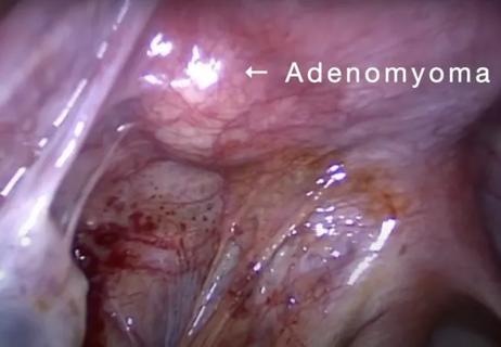
Laparoscopic surgery provides relief for teenage patient with adenomyoma and endometriosis

New research highlights serious risks and the critical need for earlier advance care planning

Initiatives focus on the physical and emotional well-being of geriatric patients and their caregivers

Researchers explore the mental and physical benefits of social prescribing

Cleveland Clinic pioneers a new paradigm in cardiovascular care delivery

Effective screening, advanced treatments can help preserve quality of life

Study suggests inconsistencies in the emergency department evaluation of geriatric patients