Virtual surgical planning and computer-aided design/computer-aided manufacturing (CAD/CAM) technology have transformed surgery to reconstruct complex maxillary and mandibular bony defects.
Advertisement
Cleveland Clinic is a non-profit academic medical center. Advertising on our site helps support our mission. We do not endorse non-Cleveland Clinic products or services. Policy
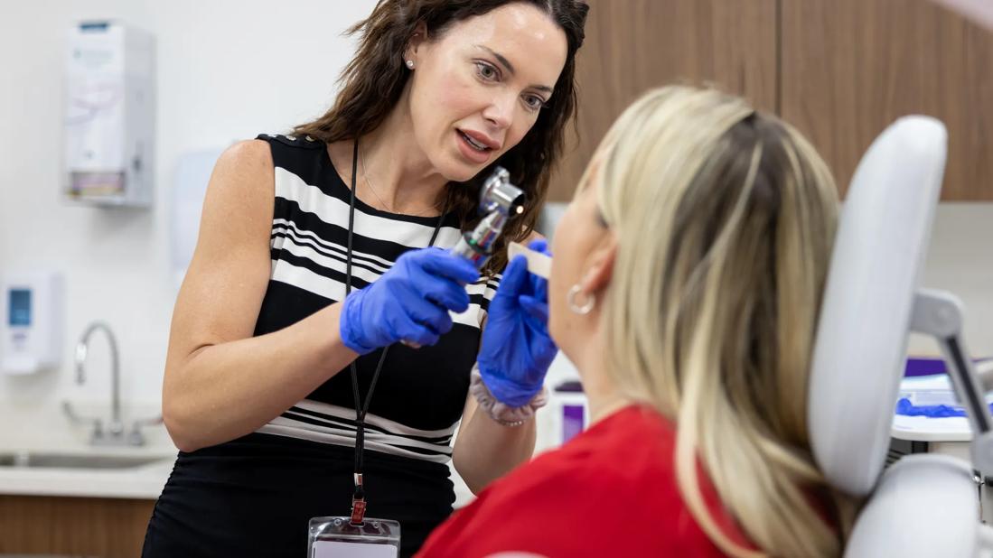
Head and neck surgeon Ashley C. Mays, MD, a recent addition to the Department of Otolaryngology at Cleveland Clinic Indian River Hospital, brings this new capability to Cleveland Clinic’s five-hospital regional health system in Florida. Currently, only a handful of centers across the state employ this advanced approach to complex head and neck bony reconstruction.
“Our goal with bony reconstruction is to restore structure and function in patients with tissue defects caused by trauma, cancer, infection or congenital deformities,” says Dr. Mays, noting malignancy is the primary origin of bony head and neck defects. “The mandible is particularly challenging to reconstruct because of its complex mobility and vital role in chewing, swallowing, speaking and facial expressions.”
She emphasizes that beyond the need to restore function so that patients can maintain independence in their activities of daily life, restoration of a natural facial appearance is necessary to improve overall quality of life and psychosocial well-being.
The gold standard for mandibular reconstruction is bony free tissue transfer, most commonly using the fibula bone. The scapula, iliac crest, and radius bones are also options when considering head and neck bony reconstruction. The fibula free flap can be used to reconstruct the bony grid of the midface and mandible with very high success rate.
“Fibula free flaps have the advantage of a reliable vascular pedicle, reduced donor site morbidity, and it provides a good base for dental implant rehabilitation,” Dr. Mays explains. “Through pairing vascularized bone with large islands of soft tissue, we can achieve reliable reconstructions for larger defects.”
Advertisement
This approach is especially useful, she notes, when patients have undergone radiation therapy, which can compromise blood supply to the area, or who have failed previous reconstruction strategies.
According to Dr. Mays, the use of virtual surgical planning and CAD/CAM software has led to a much more personalized approach to head and neck bony reconstruction. “This technology allows me to create a strategic plan based on the patient’s own scans prior to the day of surgery, taking some of the guesswork out of creating a pre-morbid fit,” she says.
Fine-cut CT scans and mirror image reconstruction data are used to virtually create three-dimensional models to plan resection and reconstruction procedures. The data are then used to manufacture patient-specific implants and surgical cutting and drilling guides. “Patient-specific implants confer a number of advantages over stock implants including better fit, strength and stability,” notes Dr. Mays.
“CAD/CAM software allows us to determine more precisely surgical margins for resections and reduces intraoperative manipulation of implants, such as plate bending,” adds Dr. Mays. “This results in greater efficiency, shorter operation times, reduced risk for complications, and more predictable results.”
At Cleveland Clinic Indian River Hospital, Dr. Mays teams with Brian Burkey, MD, MEd, Chair of the Department of Otolaryngology-Head and Neck Surgery, to care for patients with head and neck cancer. Together they use CAD/CAM technology to plan and perform ablative and reconstructive procedures.
Advertisement
“This two-surgeon approach allows us to perform tumor resection and free flap reconstruction during one surgery, which can improve functional and esthetic results,” observes Dr. Mays, who is fellowship-trained in head and neck surgical oncology and total body microvascular reconstruction.
Advertisement
Advertisement

Nonthermal technique reduces bleeding and perforation risk

Standardizing a minimally invasive approach for Barrett’s Esophagus and Esophageal Cancer

PSMA-targeted therapy for metastatic prostate cancer now offered at Cleveland Clinic Weston Hospital

Nationally recognized urologic oncologist offers vision for growth, innovation, and excellence

Noninvasive modality gains ground in United States for patients with early-to-moderate disease

Cleveland Clinic Weston Hospital’s collaborative model elevates care for complex lung diseases

Interventional pulmonologists at Cleveland Clinic Indian River Hospital use robotic technology to reach small peripheral lung nodules

Trained in the use of multiple focal therapies for prostate cancer, Dr. Jamil Syed recommends HIFU for certain patients with intermediate-risk prostate cancer, especially individuals with small, well-defined tumors localized to the lateral and posterior regions of the gland.