Locations:

Cleveland Clinic Florida performing fewer percutaneous lung biopsies
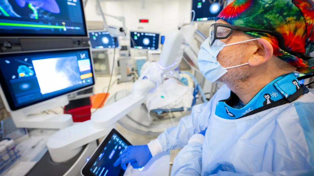
Image content: This image is available to view online.
View image online (https://assets.clevelandclinic.org/transform/fbefb16a-1d04-48ac-ab35-59c3cba101f3/PUL_5529311_02-18-25_0008_MK)
AlShelli_Ihab_Robotic Bronchoscopy
Robotic-assisted bronchoscopy (RAB) has made it possible to reach and biopsy more pulmonary nodules than ever before, a critical step for the definitive diagnosis of lung cancer, the leading cause of cancer death in the United States.
Advertisement
Cleveland Clinic is a non-profit academic medical center. Advertising on our site helps support our mission. We do not endorse non-Cleveland Clinic products or services. Policy
“Size and location are the limiting factors in choosing when and how to biopsy nodules, especially when you consider an estimated 70% are found in the outer third of the lung, beyond the ability of bronchoscopy to visualize,” says Ihab AlShelli, MD, MBChB, a pulmonary medicine specialist with Cleveland Clinic Weston Hospital. “Innovative technology is quickly changing diagnostic protocols at Cleveland Clinic Florida and has shifted thinking on when nodules may merit biopsy.”
The last 20 years have seen major advancements in lung cancer diagnostics from a pulmonary stand point, including endobronchial ultrasound (EBUS), ultrathin bronchoscopy, virtual bronchoscopy, and electromagnetic navigation bronchoscopy (ENB). “None bring as much promise as CT-guided needle biopsy performed by radiologists for the diagnosis of peripheral nodules until recently with the robotic-assisted bronchoscopy,” asserts Dr. AlShelli.
According to a recent observational study, nearly half (46%) of patients experienced multiple biopsies prior to a lung cancer diagnosis, 41% of which were bronchoscopic, 52% percutaneous, and 7% surgical. Dr. AlShelli anticipates a shift toward RAB for accessing and diagnosing peripheral nodules as more centers invest in new technology that can reach all 18 segments of the lung and lesions outside of the airway. “The introduction of robotic-assisted bronchoscopy is changing our surveillance practices and resulting in fewer CT-guided biopsies at Cleveland Clinic Florida,” he confirms.
Advertisement
The robotic system used at Cleveland Clinic Florida has a 3.5 mm outer-diameter catheter with a 2.0 mm working channel that can pass through small airways and reach to the periphery of the lung. According to Dr. AlShelli, the robotically-held catheter imparts greater stability and leaves the physician’s hands free to manipulate the finely controlled instruments used for biopsy.
“The catheter is flexible and has a removable vision probe that allows me to aim toward the target and make adjustments to the catheter’s positioning before taking tissue samples,” he describes. “We can more confidently biopsy suspicious peripheral nodules and avoid sensitive structures.”
He also points to the lower complications rates observed with bronchoscopy. The aforementioned observational study found pneumothorax was recorded in 10.2% of percutaneous patients and just 1.84% of bronchoscopy patients. While percutaneous and bronchoscopic biopsies had a similar risk of hemorrhage — occurring in 0.7% and 0.6% cases, respectively — this complication was reported in 3.3% of patients undergoing a surgical biopsy.
Increased diagnostic yields, ranging from 78-93%, is one of the major advantages of RAB. This is achieved in part by the robotic system’s ability to minimize CT to body divergence through enhanced visualization. The system’s software combines multiple imaging modalities in a single heads-up display, including radial EBUS, fluoroscopy, and even cone beam CT, allowing greater accuracy of catheter placement.
Advertisement
“These imaging modalities help cut down on the small differences that can exist between our virtual target and the actual target, which is incredibly important when 1 to 2 mm can make a big difference in yield,” says Dr. AlShelli. “Poor yields can delay a confirmed diagnosis and have a significant impact on disease progression.”
An estimated 1.6 million pulmonary nodules are detected each year in the United States as incidental findings on chest radiographs and CT scans. Although active surveillance is the standard of care for the majority as most of these nodules are benign, any nodule larger than 7 or 8 mm with intermediate to high pretest probability may merit biopsy, notes Dr. AlShelli.
“Most physicians who deal with lung nodules would wait for highly suspicious nodules to grow big enough, but with robotic-assisted biopsy it is not worth the risk,” he adds. “The right diagnostic modality can help ensure patients receive lifesaving therapy when they need it or avoid aggressive treatment when simpler measures may be enough.”
More than 14 million people are now eligible for lung cancer screening thanks to expanded guidelines issued last year from the US Preventive Services Task Force. While only 6.5% currently take this critical step each year, Dr. AlShelli is optimistic more Americans will seek lung cancer screening due to broader Medicare coverage announced this year and a requirement for most private insurance plans to cover screening for those at a high risk.
“As screening rates improve, we are sure to see an increase in the identification of small, hard to reach pulmonary nodules,” says Dr. AlShelli. “Today, with robotic-assisted bronchoscopy, we can offer a better approach to achieve a definitive diagnosis.”
Advertisement
Advertisement

Study highlights strong predictive value of circulating tumor DNA testing

Researchers with Cleveland Clinic in Florida highlight need for a national registry for gastrointestinal stromal tumors

Multidisciplinary LARS Center addresses life-altering consequence of rectal cancer surgery

Early recognition and intervention recommended in cubital tunnel syndrome

Manpreet (Meena) Bedi, MD, named Division Chair of Radiation Oncology

AR-assisted navigation is closing the gap between surgical planning and implant placement

Evidence shows early evaluation improves survival and quality of life – yet many eligible patients are referred too late

2026 ADA Standards of Care promote holistic, multisystem management