Locations:

Cole Eye Institute imaging specialists are equal parts technician, artist and diagnostician
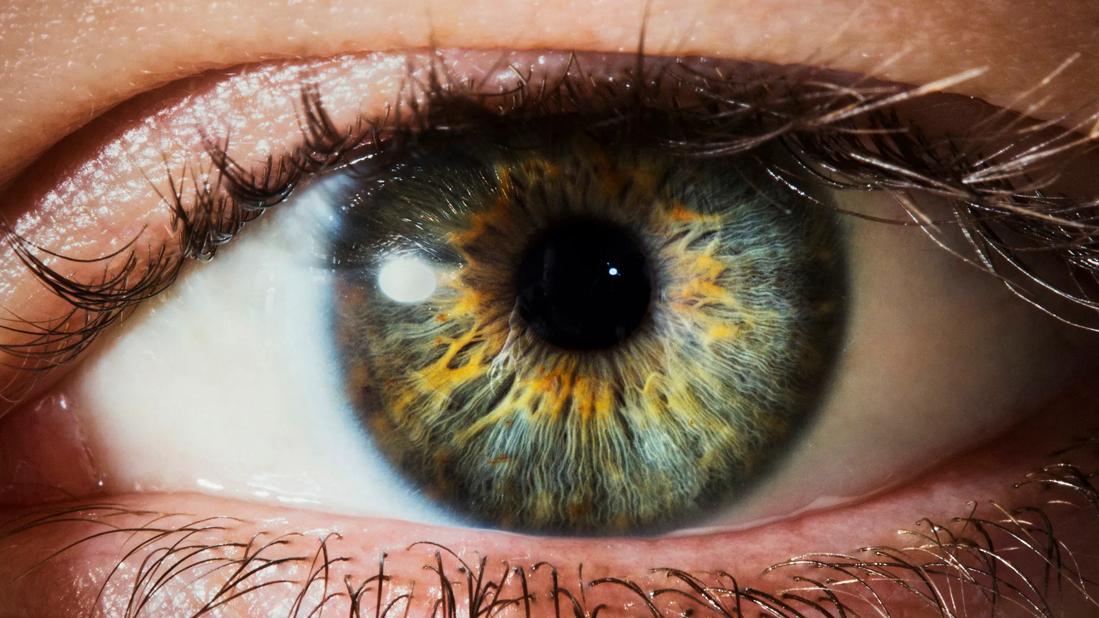
Image content: This image is available to view online.
View image online (https://assets.clevelandclinic.org/transform/74fdb2c6-7781-43b3-bd16-1117e56a3263/eyeFacts-146599805-770x533-1-jpg)
Eye close-up
Part technician, artist and diagnostician, ophthalmic imaging specialists have risen in prominence at Cleveland Clinic Cole Eye Institute. In the 1990s, the percentage of Cole Eye Institute patients requiring imaging was about 5%. Today, it’s more than 50% — which explains why Cole Eye Institute accounts for the most clinical images captured at Cleveland Clinic outside of the Imaging Institute. The number of eye images is on track to total nearly 180,000 in 2023.
Advertisement
Cleveland Clinic is a non-profit academic medical center. Advertising on our site helps support our mission. We do not endorse non-Cleveland Clinic products or services. Policy
Ophthalmic imaging — including anterior- and posterior-segment photography, fluorescein and indocyanine green angiography, B-scan ultrasound, and ultrasound biomicroscopy — has become essential for diagnosis in modern ophthalmology. That’s the reason imaging specialists will be integrated with care teams in the new Cole Eye Institute building, moved out of their designated space in the current building to be closer to patients.
“We used to be called ‘ophthalmic photographers,’” says Brandy H. Lorek, one of the 14 imaging specialists at the Cole Eye Institute and manager of the department. “Now, many of us do more than photography and have certifications in different types of imaging.”
It’s not a well-marked (or widely traveled) career path, she says. Few universities offer degrees in ophthalmic imaging. Most specialists fall into the career unexpectedly and learn on the job, with disciplines and techniques passed on similar to an apprenticeship.
After earning a bachelor’s degree in biology, Lorek worked in an ocular oncology lab at Bascom Palmer Eye Institute. A clinician researcher there suggested she pursue mastering ophthalmic ultrasound. She began studying under another ophthalmic echographer and became certified by the International Joint Commission on Allied Health Personnel in Ophthalmology.
“There’s no school or certification for most ophthalmic imaging skills,” she notes. “You need to learn the practices from someone else.”
Using the imaging instrument is only half the job. The other half is understanding disease processes.
Advertisement
“Ophthalmic imaging specialists pay extremely close attention to detail,” says Lorek. “We know what to look for in certain eye diseases, and we do imaging with that knowledge. That’s what separates us from someone just taking pictures. We know what ophthalmologists are trying to see, so we know what to capture to help them diagnose or rule out a disease.”
The more academic knowledge you have in the field of ophthalmology, the better imaging you can capture.
“I can teach someone to use a camera in one day, but that doesn’t make them an imaging specialist,” says Lorek. “It’s easy to image the back of an eye but miss all the pathology.”
Expert imaging is especially important at the Cole Eye Institute, where medical teams routinely encounter patients with severe, advanced, rare or even unique eye conditions.
“Patients travel to us from out of state for diagnoses that sometimes require a high-quality fluorescein angiogram or other specialized imaging,” says Lorek. “We’ve seen some amazing things.”
Below is a collection of notable images captured by Cole Eye Institute ophthalmic imaging specialists.
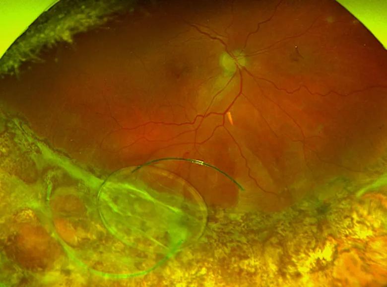
Image content: This image is available to view online.
View image online (https://assets.clevelandclinic.org/transform/d2349dae-7c83-4b3f-987f-6528ef3baae4/23-EYE-3994158-CQD-Remarkable-Eye-Images-Inset-1_jpg)
This ultra-widefield fundus image taken by certified ophthalmic assistant (COA) Amy Forman depicts a dislocated intraocular lens resting on the surface of the retina. This eye had a history of retinal detachment and scleral buckle surgery, as evident by the white matter at the bottom of the image.
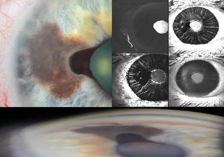
Image content: This image is available to view online.
View image online (https://assets.clevelandclinic.org/transform/2871dcf2-6b07-4620-bdd7-3905992f82b8/23-EYE-3994158-CQD-Remarkable-Eye-Images-Inset-2_jpg)
This composite image of an iris melanoma was taken by Mark Harrod, a certified retinal angiographer (CRA) who also is certified in optical coherence tomography (OCT-C). It includes modalities such as slit-lamp and gonio imaging, and anterior-segment fluorescein angiography, highlighting the lesion’s thickness and internal vascularity. This composite was among those awarded at the American Academy of Ophthalmology (AAO) 2019 meeting, as part of the Ophthalmic Photographers’ Society annual contest.
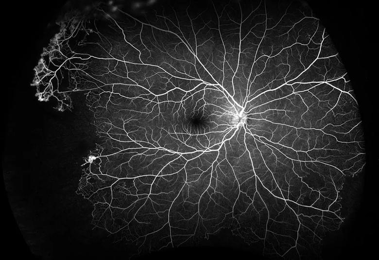
Image content: This image is available to view online.
View image online (https://assets.clevelandclinic.org/transform/0af183f6-97af-4e55-896f-92806e5ccddf/23-EYE-3994158-CQD-Remarkable-Eye-Images-Inset-3_jpg)
Another 2019 Ophthalmic Photographers’ Society award-winner, this fluorescein angiogram by Mark Harrod, CRA, OCT-C, depicts peripheral (sea fan) neovascularization and peripheral nonperfusion in a patient with sickle cell disease.
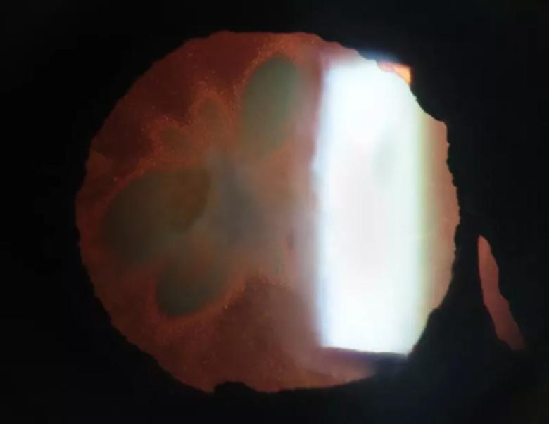
Image content: This image is available to view online.
View image online (https://assets.clevelandclinic.org/transform/163e07ae-16d6-433d-ae6f-3dc3c938dd2c/23-EYE-3994158-CQD-Remarkable-Eye-Images-Inset-4_jpg)
Lisa Jahn-Gregory, OCT-C, COA, captured this retro-illuminated slit-lamp image of a cataract in the shape of a butterfly.
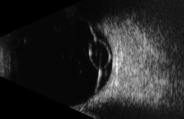
Image content: This image is available to view online.
View image online (https://assets.clevelandclinic.org/transform/48fda4f3-89cb-4178-9764-fa58ab0e8fd1/23-EYE-3994158-CQD-Remarkable-Eye-Images-Inset-5_jpg)
This ultrasound image reveals a retinal detachment with a large retinal cyst, indicative of a chronic retinal detachment. The image was captured by Beth MacQueen, a certified diagnostic ophthalmic sonographer (CDOS) and a registered ophthalmic ultrasound biometrist (ROUB).
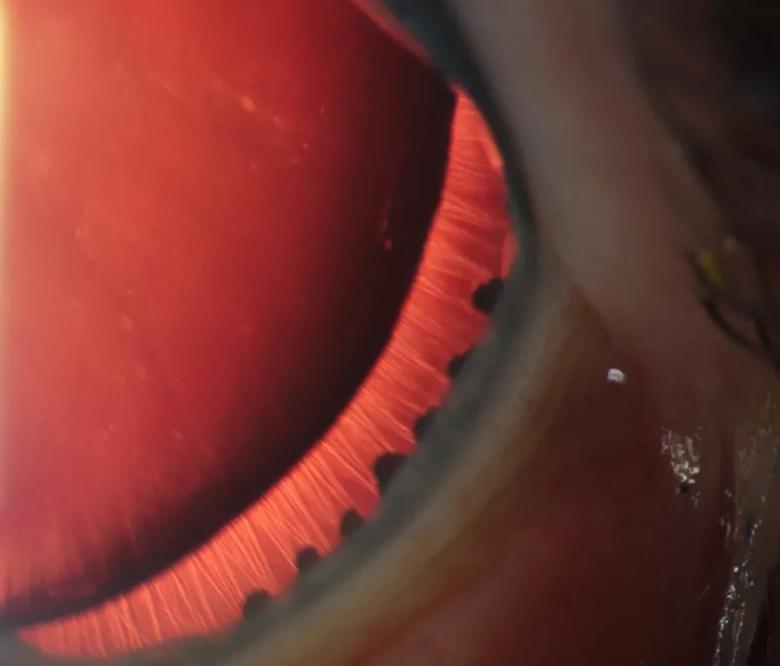
Image content: This image is available to view online.
View image online (https://assets.clevelandclinic.org/transform/c5029bf1-5648-44b7-8a4e-ac2004757321/23-EYE-3994158-CQD-Remarkable-Eye-Images-Inset-6_jpg)
Certified ophthalmic technician (COT) Stephanie Pelton, OCT-C, took this retro-illuminated slit-lamp image of a patient with aniridia (absence of the iris). This image shows all the zonules that connect the ciliary processes to the periphery of the lens.
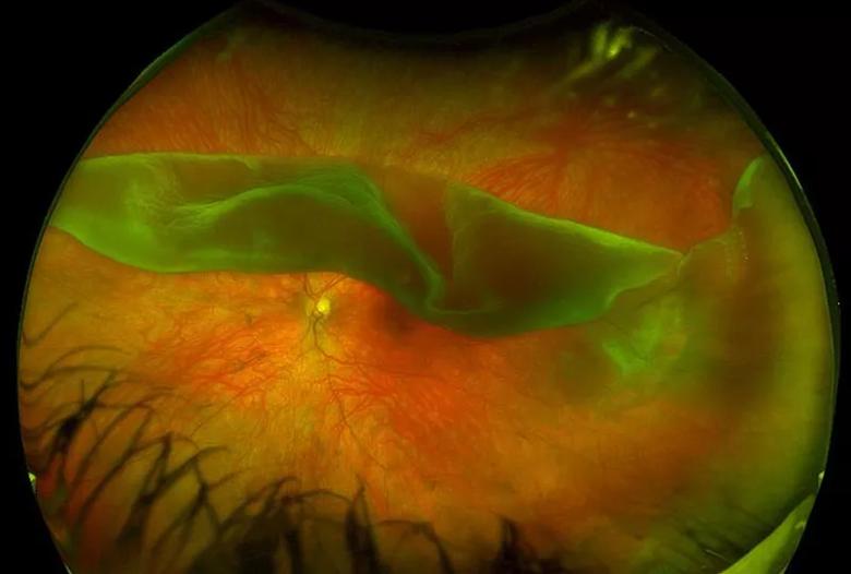
Image content: This image is available to view online.
View image online (https://assets.clevelandclinic.org/transform/f808a880-209c-490f-8260-c34615da566d/23-EYE-3994158-CQD-Remarkable-Eye-Images-Inset-7_jpg)
This ultra-widefield fundus image, captured by Lindsey VanAuker, OCT-C, COA, reveals a giant retinal tear obscuring the patient’s central vision.

Image content: This image is available to view online.
View image online (https://assets.clevelandclinic.org/transform/59a295f8-4e5f-47ce-82a8-3a9dc0302e04/23-EYE-3994158-CQD-Remarkable-Eye-Images-Inset-8_jpg)
This ultrasound biomicroscopy image by Brandy H. Lorek, CDOS, ROUB, shows a grossly centered presbyopia-correcting intraocular lens with dislocated haptics, causing uveitis glaucoma hyphema (UGH) syndrome.
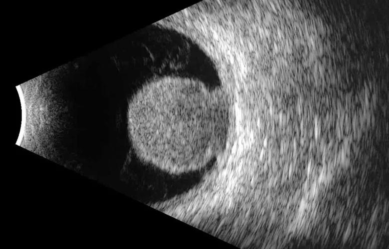
Image content: This image is available to view online.
View image online (https://assets.clevelandclinic.org/transform/a0bc31b7-3c7d-4f15-86cf-8fa88e72d3df/23-EYE-3994158-CQD-Remarkable-Eye-Images-Inset-9_jpg)
A large choroidal melanoma was captured on ultrasound by Brandy H. Lorek, CDOS, ROUB. The tumor’s rupture of Bruch’s membrane resulted in the pathognomonic collar-button shape.
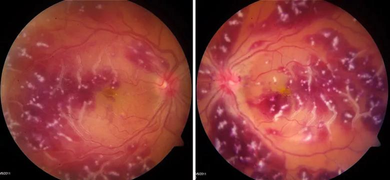
Image content: This image is available to view online.
View image online (https://assets.clevelandclinic.org/transform/4b580e05-28e6-43df-86d8-40b6fd437658/23-EYE-3994158-CQD-Remarkable-Eye-Images-Inset-10_jpg)
These fundus images illustrate severe retinopathy and vasculitis, a rare ophthalmic manifestation of dermatomyositis.
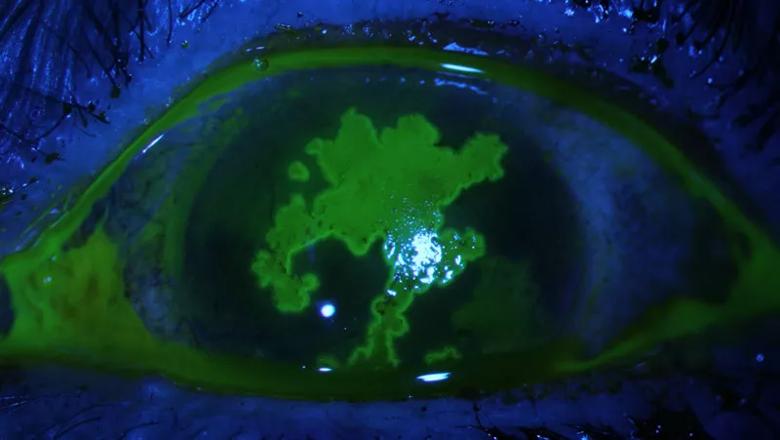
Image content: This image is available to view online.
View image online (https://assets.clevelandclinic.org/transform/06e67b4b-291b-4491-97cc-fb63d150e61e/23-EYE-3994158-CQD-Remarkable-Eye-Images-Inset-11_jpg)
An epithelial defect secondary to herpes simplex virus is depicted in this slit-lamp image using fluorescein stain. This large, well-defined defect caused severe pain and vision impairment.
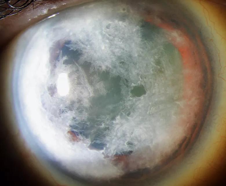
Image content: This image is available to view online.
View image online (https://assets.clevelandclinic.org/transform/b481dbaf-b521-44b7-81b7-1f921908fc54/23-EYE-3994158-CQD-Remarkable-Eye-Images-Inset-12_jpg)
This slit-lamp image with broad-beam lighting shows diffuse infiltration of the cornea in a patient with multiple myeloma.

Image content: This image is available to view online.
View image online (https://assets.clevelandclinic.org/transform/23740085-e095-4537-8d32-1615b9e987c3/23-EYE-3994158-CQD-Remarkable-Eye-Images-Inset-14_jpg)
This early frame from a fluorescein angiogram was captured with an ultra-widefield fundus camera by Stephanie Burke, CRA, OCT-C. This image illustrates retinal manifestations of Wyburn-Mason syndrome, including multiple arteriovenous malformations.
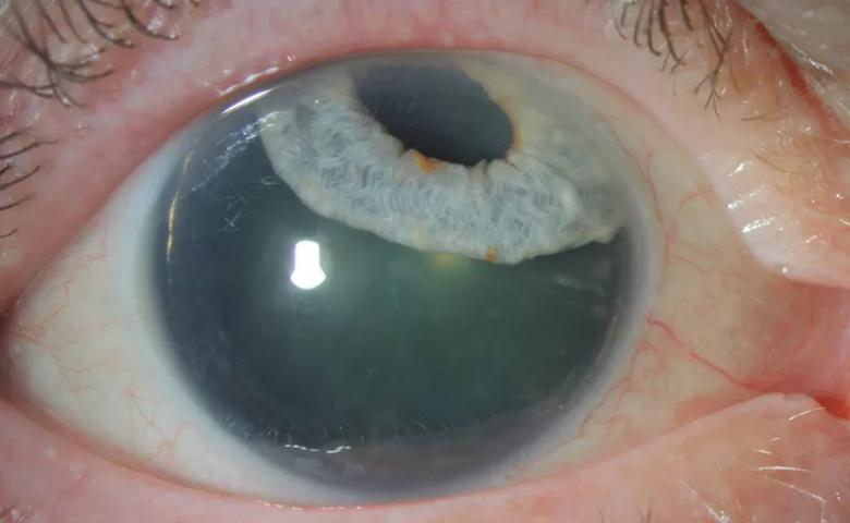
Image content: This image is available to view online.
View image online (https://assets.clevelandclinic.org/transform/4961798f-1a9e-4f4e-bab1-97dc4af47ec5/23-EYE-3994158-CQD-Remarkable-Eye-Images-Inset-13_jpg)
This slit-lamp photograph captured by Stephanie Burke, CRA, OCT-C, depicts iridodialysis (disinserted iris) secondary to severe trauma. This image shows the patient looking straight at the camera.
Advertisement
Advertisement

Imaging dye enables vascular assessment to promote procedural precision and safety

The new application that could augment cancer detection, improve efficiency

How computational tools and personalized biomechanics can improve keratoconus detection, ectasia risk assessment and surgical outcomes

Two studies from Cleveland Clinic may help advance the technology toward broader clinical use

Overcoming barriers to implementing clinical trials

Multimodal evaluations reveal more anatomic details to inform treatment

A closer look at the impact on procedures and patient outcomes

Join us in Florida this winter for a long-standing CME favorite