Novel surgical approach provides better outcomes for patients

By Erika Woodson, MD, FACS
Advertisement
Cleveland Clinic is a non-profit academic medical center. Advertising on our site helps support our mission. We do not endorse non-Cleveland Clinic products or services. Policy
Endoscopic ear surgery (EES) has emerged as a major advancement in the otologist’s chronic ear disease armamentarium. EES enables improved surgical visualization, allowing for better elimination of disease, improvement of aeration pathways and reduction of open (postauricular) approaches.
Postauricular approaches have been standard for many middle ear pathologies, but EES is changing this. Both the visualization and instrumentation have matured so that the endoscopic otologist can avoid a postauricular approach in many patients — including children. As a result, the patient is spared the additional pain and healing time required of a postauricular incision.
Ear surgeons at Cleveland Clinic have been incorporating endoscopes into otologic surgery for nine years and will continue to explore the limits of this technique. EES is particularly useful in cases of cholesteatoma, a type of noncancerous skin growth that forms beyond the normal boundary of the eardrum. It causes chronic infection, ear drainage and destruction of the hearing bones and mastoid (skull around the ear). Removal of this abnormal skin is the only treatment option. Complete resection and proper ventilation are critical to preventing recurrence, ongoing infection or progression of bony destruction.
Cholesteatoma is difficult to access with traditional microscopic approaches. In traditional microscopic ear surgery, the surgical view is limited by what is directly visible with an external
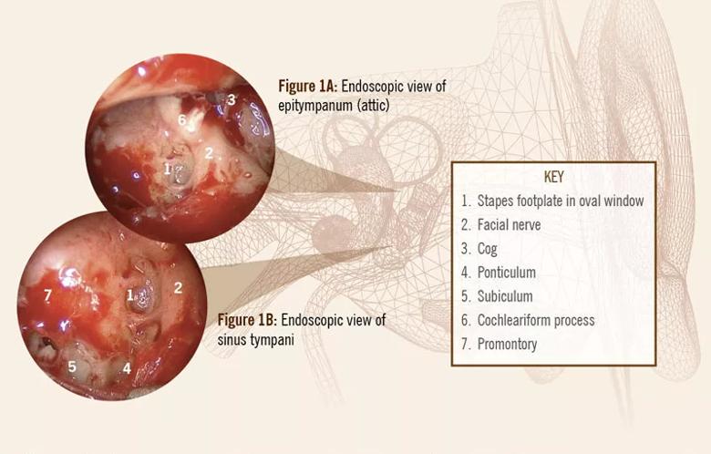
microscope’s light beam. In contrast, EES allows for superior inspection of structures that are otherwise hidden and are frequently invaded by cholesteatoma, namely the anterior mesotympanum, epitympanum (attic) (Figure 1A) and sinus tympani (Figure 1B).
Advertisement

Image content: This image is available to view online.
View image online (https://assets.clevelandclinic.org/transform/e34b33cc-e206-4c74-b9e8-0c61713c093c/19-ENT-4249-endocopicViews1-inset-800x511_jpg)
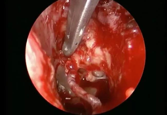
In another case illustration, a surgical endoscope was used to remove a locally advanced cholesteatoma. Because of the increased visualization of the 30-degree endoscope (see video below), we were able to eradicate the cholesteatoma without sacrificing the ossicles. Techniques like this stand to improve hearing and control rates of cholesteatomas for chronic ear disease patients.
Video content: This video is available to watch online.
View video online (https://www.youtube.com/embed/9p-dPLQ1r8I?feature=oembed)
Surgical endoscope to remove a locally advanced cholesteatoma
The decision to use EES versus a microscopic approach is an important one. EES may not be feasible or recommendable in all cases, nor does it mean necessarily shorter surgeries. However, endoscopes provide greater visualization than microscopes. In experienced hands, this can mean a more thorough surgery with fewer incisions. These benefits have immediate implications for surgical recovery and long-term implications for the surgical correction of chronic ear disease.
Dr. Woodson is Section Head, Otology-Neurotology and Medical Director of the Hearing Implant Program in Cleveland Clinic’s Head & Neck Institute.
Advertisement
Advertisement

Analysis of HNSCC patients shows HPV status to be predictive of higher abundance of oncobacteria within the tumor

Case study illustrates the potential of a dual-subspecialist approach

Evidence-based recommendations for balancing cancer control with quality of life

Study shows no negative impact for individuals with better contralateral ear performance

HNS device offers new solution for those struggling with CPAP

Patient with cerebral palsy undergoes life-saving tumor resection

Specialists are increasingly relying on otolaryngologists for evaluation and treatment of the complex condition

Detailed surgical process uncovers extensive middle ear damage causing severe pain and pressure.