Locations:

Five categories of cutaneous abnormalities are associated with COVID-19
By Samantha Polly, MD, and Anthony P. Fernandez, MD, PhD
Advertisement
Cleveland Clinic is a non-profit academic medical center. Advertising on our site helps support our mission. We do not endorse non-Cleveland Clinic products or services. Policy
This article has been excerpted and reprinted (without references) from the Cleveland Clinic Journal of Medicine (2022, 89(3):161-167. The open-access and fully referenced original article is available here.
As experience with caring for patients with COVID-19 has accumulated since the onset of the pandemic, so has our understanding of its associated cutaneous manifestations and their clinical implications.
It is beneficial to watch for cutaneous manifestations of COVID-19, both in and out of the hospital. For example, a study of more than 330,000 community-based patients in the United Kingdom found that patient-reported skin rash was associated with positive COVID-19 testing and was more predictive than fever. Additionally, an analysis of 296 hospitalized patients with COVID-19 in the United States found that mucocutaneous findings were associated with the need for mechanical ventilation, even when adjusted for age, body mass index and comorbidities.
COVID-19-associated cutaneous abnormalities are often grouped into five major categories:

Image content: This image is available to view online.
View image online (https://assets.clevelandclinic.org/transform/19c0b76f-fd3b-4f38-a150-3c446e4368a6/22_PSX-3046994_CQD-Hero-6540x450-1_jpg)
Morbilliform eruptions are common in many viral illnesses and were reported in patients with COVID-19 early in the pandemic. International registry data indicate that morbilliform eruptions are the most common cutaneous manifestation in patients with laboratory-confirmed COVID-19. Typical clinical features include a generalized, symmetric maculopapular rash with pruritus.
Advertisement
Patients with COVID-19-associated morbilliform eruptions have an excellent prognosis, with survival rates of 96.9% to 97.5%.
Urticaria is also common in COVID-19. The clinical features do not appear to differ from those of idiopathic urticaria and typically consist of generalized pruritic wheals. On average, urticaria lasts less than one week and is associated with relatively mild disease and survival rates of 97.8% to 98.2%.
Histologic features also mimic those of idiopathic urticaria and thus limit the value of skin biopsy. However, urticarial vasculitis has been described in association with COVID-19, suggesting that biopsy should be considered in patients with persistent urticarial plaques with associated purpura.
Interestingly, in a systematic review of 895 patients with COVID-19, 105 (12%) had urticarial lesions, and in 17 (16%) of these 105 the urticaria began before the onset of the other COVID-19 symptoms, suggesting that it can be a clue to diagnosis in appropriate clinical settings and can help guide early testing. Additionally, an analysis of 200 patients with COVID-19 with cutaneous manifestations found a significant association between urticarial and gastrointestinal symptoms, which could assist clinicians in their anticipatory management.
Initially described as “varicella-like,” vesicular eruptions in COVID-19 have been described in both localized and diffuse distributions. The localized pattern is characterized by monomorphic vesicles in the same stage of evolution that are confined to the trunk. But the diffuse pattern may be more common. A cohort study reported that it accounted for 18 (75%) of 24 cases. The diffuse pattern consists of polymorphic papules, vesicles, and pustules that develop simultaneously on the trunk and spread distally, sometimes involving the palms and soles. Lesions tend to resolve after about eight days without scarring.
Advertisement
Lesional skin biopsies reveal histologic features consistent with viral exanthems, namely vacuolar gegeneration of the basal epidermal layer with occasional dyskeratotic keratinocytes and superficial dermal inflammation. However, some reports describe prominent keratinocyte acantholysis contributing to formation of intraepidermal vesicles, which is a relatively unusual histologic finding.
Additionally, while there have been reports of SARS-CoV-2 spike proteins detected with immunohistochemistry in sweat glands and dermal endothelial cells in skin biopsies from COVID-19 patients, studies of COVID-19-associated vesicular rashes detected no SARS-CoV-2 in vesicular fluid by reverse transcriptase polymerase chain reaction testing.
Like urticaria, vesicular eruptions were also commonly noted before other COVID-19 symptoms (in 8.5% to 15% of cases of COVID-19-associated urticaria) in multiple studies, and therefore may similarly provide an indication for COVID-19 testing and isolation in the appropriate clinical context. Additionally, a systematic review reported a possible link between vesicular eruptions and neurologic symptoms, including headache, dysgeusia, irritability and confusion. Like those with morbilliform rash or urticaria, patients with COVID-19 with vesicular eruptions have high survival rates (96.1% to 96.6%).
Although pernio-like acral lesions were the first cutaneous manifestations to generate significant attention, whether they are truly linked to COVID-19 has been debated.
Advertisement
Challenging the link are strikingly low rates of positive COVID-19 testing in affected patients, as well as results of several studies that suggest these lesions are most consistent with typical perniosis, with an increased incidence related to changes in daily routine (such as quarantining and working from home) during the pandemic rather than infection with SARS-CoV-2. Additionally, a systematic review found that pre-existing rheumatologic conditions were more common in patients with presumed COVID-19-related pernio-like lesions, raising the possibility that underlying diagnoses contributed to development of the acral lesions.
However, proponents of the association with COVID-19 point to outbreaks of chilblain-like lesions corresponding to COVID-19 waves and propose that an efficient, type I interferon-driven antiviral response could induce pernio-like lesions and suppress both symptoms and confirmatory testing.
Interestingly, information is accumulating about pernio-like lesions in long-hauler patients, with a significant association reported between persistent cutaneous and extracutaneous symptoms.
While the debate continues, if these lesions are truly a COVID-19 manifestation, they are, fortunately, associated with high survival rates (96.4% to 98.7%) and few or no systemic symptoms.
Vaso-occlusive lesions have been reported in patients with COVID-19 with varied clinical presentations, including fixed livedo racemosa, retiform purpura, and acral ischemia, which may be clinically confused with COVID toes. These lesions are most commonly seen in hospitalized patients with moderate to severe COVID-19 and are associated with higher risks of severe pneumonia and intensive care unit admission and relatively low survival rates (78.9% to 81.8%). Similar patterns of microvascular thrombosis have been found in skin biopsies and pulmonary tissue of COVID-19 patients with vaso-occlusive cutaneous lesions, suggesting that this manifestation could be a marker of systemic microvascular injury. Additionally, systemic thrombotic events including deep vein thrombosis and pulmonary embolism have been reported in patients with retiform and necrotic lesions, with rates as high as 64%. Whether early recognition of these lesions can prompt treatment decisions that decrease systemic thrombotic events or increase overall survival requires further research.
Advertisement
Other cutaneous findings that have been reported with COVID-19 include oral lesions; reactivation of viral infections; rash resembling symmetrical drug-related intertriginous and flexural exanthema; small-vessel vasculitis; cutaneous hyperesthesia; papulosquamous eruptions; and erythema nodosumlike lesions.
Oral lesions. A study of 666 patients reported various oral mucosal findings in 78 (26%) of 304 patients who had mucocutaneous manifestations, and the authors hypothesized that lesions in the mouth may be under reported due to contact precautions and assisted ventilation that limits examination of the oral mucosa.
Herpes simplex virus (HSV) and varicella-zoster virus (VZV) reactivations have been reported in conjunction with COVID-19 infection. A cross-sectional study of nearly 900 patients with COVID-19 found a significantly higher prevalence of HSV-1 and VZV than in the hospital population, even when adjustments were made for numerous comorbidities. Some reports suggest that HSV reactivation may be associated with more severe COVID-19 infection, including acute respiratory distress syndrome and viremia, but the prognostic implications of treating these reactivations has not yet been robustly investigated.
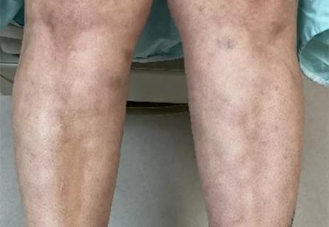
Image at top shows vaso-occlusive lesions in COVID-19. Left, a 62-year-old man with COVID-19 developed an irregular, mottled, purpuric patch on his knee extending onto his thigh during an extended hospitalization complicated by septic shock and acute respiratory failure requiring mechanical ventilation. He died of his illness 3.5 weeks after admission. At right, a 77-year-old man developed purpuric patches with central hemorrhagic crusts on the left buttock shortly after hospitalization for COVID-19.
Advertisement
Patients report improved sense of smell and taste
Clinicians who are accustomed to uncertainty can do well by patients

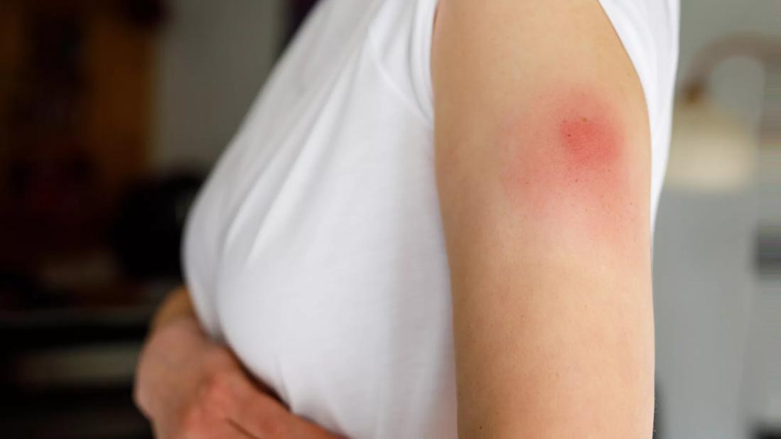
Unique skin changes can occur after infection or vaccine

Cleveland Clinic analysis suggests that obtaining care for the virus might reveal a previously undiagnosed condition

As the pandemic evolves, rheumatologists must continue to be mindful of most vulnerable patients
Early results suggest positive outcomes from COVID-19 PrEP treatment
Could the virus have caused the condition or triggered previously undiagnosed disease?

Black race, COVID-19 hospitalization and mood symptoms confer increased risk, study finds