Locations:

Sinus tracts can occur years later and not near the incision site
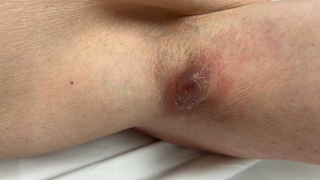
Image content: This image is available to view online.
View image online (https://assets.clevelandclinic.org/transform/d7c47b9a-1773-4575-ae9c-dd46c861d644/shoulder-periprosthetic-joint-infection3)
An open wound in a patient's armpit
At age 66, a patient had shoulder hemiarthroplasty for a proximal humerus fracture. Seventeen years later, he presented with a draining wound in his axilla. He was evaluated by a dermatologist and underwent local wound care for several months. Eventually he was referred to a shoulder specialist, who diagnosed him with a periprosthetic joint infection (PJI) caused by common bacteria Cutibacterium acnes (C. acnes). His atypical presentation of infection after shoulder arthroplasty was treated with an excision of the sinus tract and a complete one-stage revision surgery, eliminating the infection.
Advertisement
Cleveland Clinic is a non-profit academic medical center. Advertising on our site helps support our mission. We do not endorse non-Cleveland Clinic products or services. Policy
This case is one of 12 atypical wound presentations of shoulder PJI recently published in a case series by a Cleveland Clinic shoulder research team.
“Shoulder PJI doesn’t always manifest in a straightforward way,” says the study’s senior author, Vahid Entezari, MD, a Cleveland Clinic shoulder and elbow surgeon. “PJI of the hip and knee usually manifests acutely as a hot, red, swollen joint with drainage of pus from the incision. PJI of the shoulder has a chronic and more indolent presentation, without typical signs of infection.”
By publishing their case series, Dr. Entezari and the research team hope to raise awareness of atypical presentations of shoulder PJI and encourage providers to more quickly refer patients with a history of shoulder arthroplasty to shoulder specialists for care.
Shoulder PJI is often caused by C. acnes or other indolent bacteria, such as methicillin-susceptible Staphylococcus aureus (MSSA) or Staphylococcus epidermidis, which live in or on the skin of the upper extremity, explains Cleveland Clinic shoulder and elbow surgeon Charles Cogan, MD, the study’s lead author. Hip and knee PJI is often caused by virulent bacteria, such as methicillin-resistant Staphylococcus aureus (MRSA).
“The indolent bacteria can be introduced in the joint during surgery,” Dr. Cogan says. “Sometimes the bacteria merely colonize a joint but don’t cause any symptoms. We aren’t certain why the bacteria sometimes become more aggressive, causing acute infection, but it may be related to the patient’s immune system. If the immune system becomes weakened or busy, it may allow an infection to emerge after being tempered for many years.”
Advertisement
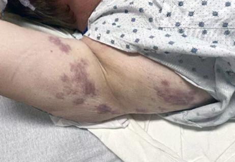
Atypical presentation of shoulder PJI may include a draining sinus tract away from the incision. This can be confusing, since the patient may have minimal symptoms in the infected joint, causing clinicians to consider a local skin or gland infection instead of PJI. In the case series:

Image content: This image is available to view online.
View image online (https://assets.clevelandclinic.org/transform/3ceede02-104f-4bdc-87ea-bd3959fc981f/shoulder-periprosthetic-joint-infection2)
A patient’s well-healed anterior shoulder incision shows no evidence of infection.

Image content: This image is available to view online.
View image online (https://assets.clevelandclinic.org/transform/d7c47b9a-1773-4575-ae9c-dd46c861d644/shoulder-periprosthetic-joint-infection3)
The same patient has a wound in the axilla, an atypical presentation of shoulder periprosthetic joint infection.

Image content: This image is available to view online.
View image online (https://assets.clevelandclinic.org/transform/af7cc4e4-ee32-48e5-a789-c1b031289a26/shoulder-periprosthetic-joint-infection1)
The patient’s X-ray indicates osteolysis around the shoulder implant, concerning for infection.
Based on the Musculoskeletal Infection Society’s criteria for PJI, infection is definite when there is a draining sinus tract or two positive tissue cultures with phenotypically identical virulent organisms. Without those major criteria, PJI still can be possible depending on other minor criteria, such as positive inflammatory markers, intraoperative cultures and implant loosening.
Diagnosis of shoulder PJI starts with a systemic collection of laboratory, clinical and radiographic data, says Dr. Entezari.
“Most often, there is stiffness, pain and radiographic changes around the implant, such as resorption of the bone and implant loosening,” he says. “However, these patients often don’t have abnormal laboratory tests, such as inflammatory markers and elevated white blood cell counts. The aspiration of the joint commonly does not yield any fluid, and in cases that aspiration is successful, the culture may be negative due to the indolent nature of the infection. Because of this, tissue cultures are kept for 14 days to increase the chance of a positive lab result.”
Advertisement
Often, an infection is not diagnosed until after a shoulder revision surgery, he adds.
“We commonly operate on patients with prior shoulder arthroplasty because of persistent pain, loss of function or mechanical failure of the implant,” he says. “We may not have any concrete evidence of infection, and culture results days or weeks after surgery make real-time intraoperative decision-making challenging.”
As a result of atypical presentation, patients have experienced delayed diagnosis of shoulder PJI. In the case series, patients presented to non-shoulder specialists (e.g., primary care, general surgery, dermatology, general orthopaedics) and commonly were referred to a wound care clinic. They received various treatments for their wounds (e.g., repeat wound debridement, vacuum-assisted wound closure), and definitive treatment was delayed for 12 months, on average.
“Besides emotional and psychological stress, delayed treatment can lead to increased complexity of subsequent surgeries, with implant loosening and progressive bone loss,” Dr. Cogan says. “This can affect the outcome of revision surgery and lead to less favorable outcomes. Especially when there’s a sinus tract and a wider area of involvement, treating the infection becomes more challenging.”
“We need to shorten the time to diagnosis for patients with shoulder PJI,” Dr. Entezari says.
Aside from quicker and more sensitive diagnostic tests (many currently in development), quicker referral to shoulder specialists can prevent a delay in care and help produce a better outcome, he says. Providers should consider referring patients to shoulder specialists earlier when an atypical wound manifests after shoulder replacement, including sinus tracts not over the incision site.
Advertisement
Shoulder PJIs occur after only 1%-2% of primary total shoulder arthroplasties, but their prevalence is growing as the number of shoulder arthroplasties increases.
“Compared to two decades ago, we are doing a staggering number of shoulder arthroplasties today,” Dr. Entezari says. “That’s because we have gotten better at recognizing shoulder problems. We now know that shoulder replacement can improve pain and function for a wider range of patients. Our indications, implant technology and postoperative care all have improved, and we know a lot more about predictors of outcome after surgery.”
To improve access to care, shoulder replacement today is often done by orthopaedic surgeons who are not shoulder specialists. But revision arthroplasty is a different beast, warns Dr. Entezari.
“I think a patient’s outcome and the result of the revision surgery may be drastically different if a shoulder specialist is engaged early in the process, especially when an infection may be involved,” he says.
Advertisement
Advertisement

Protein expression in synovial fluid indicates patients’ immune factors may be involved

Collaboration must cross borders and disciplines

Clinical complexity demands personalized care strategies

Higher than 5-year rates for prostate cancer and melanoma and about the same as breast cancer
Researchers hope it may one day help patients avoid explantation surgery
Symptoms complement one another

Innovative procedure offers less-invasive alternative to Latarjet procedure

Even patients with reported penicillin allergies can receive it without increased complications