Paired blood and brain tissue methylation findings raise prospect of noninvasive precision diagnosis

Image content: This image is available to view online.
View image online (https://assets.clevelandclinic.org/transform/54554830-c49e-4071-912a-346b9ea22c37/methylation-biomarkers-in-focal-cortical-dysplasia-135018352)
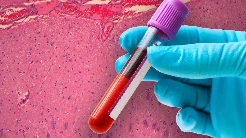
gloved hand holding blood vial with neuropathology slide in background
Researchers have identified and validated three specific DNA methylation biomarkers capable of distinguishing subtypes of focal cortical dysplasia (FCD), a common cause of drug-resistant epilepsy. Notably, these epigenetic markers (IL1RAP, HIPK2 and CNMD) were detectable not only in resected brain tissue but also in matched peripheral blood samples from the same patients.
Advertisement
Cleveland Clinic is a non-profit academic medical center. Advertising on our site helps support our mission. We do not endorse non-Cleveland Clinic products or services. Policy
The discovery, published in Brain Communications (2025;7[4]:fcaf277) by investigators from Cleveland Clinic and the Baker Heart and Diabetes Institute in Australia, opens the door to a potential noninvasive blood test for presurgical diagnosis of FCD. This would represent a significant leap forward for patient management, particularly in cases where FCD lesions are subtle or not visible on standard MRI.
“This study shows that epigenetic markers in blood can reflect changes in brain tissue in FCD subtypes IIa and IIb,” says co-author Imad Najm, MD, Director of Cleveland Clinic’s Epilepsy Center. “For clinicians, this may mean earlier and more accurate diagnostic stratification and more-personalized care. For patients, it offers the hope of less-invasive testing and ultimately better treatment outcomes.”
FCD represents a spectrum of brain malformations that originate during embryonic development and are a leading cause of drug-resistant epilepsy in adults and children. Despite advancements in neuroimaging and neuropathology, classification of FCDs has remained challenging in view of their intricate nature and often subtle architectural disorganization, which can make them difficult to detect even with the use of highest-resolution MRI. This diagnostic ambiguity can complicate decision-making around epilepsy surgery and even rule out some patients from being considered for surgery, impacting treatment outcomes.
The latest International League Against Epilepsy classification of FCDs used a tiered system that integrates genetic and epigenetic findings with clinical and imaging data. Prior research has highlighted the role of germline and somatic mutations in FCD IIa and IIb.
Advertisement
Beyond genetic sequence alterations, epigenetic modifications, such as DNA methylation, are key to regulating gene expression without changing the underlying DNA sequence. Prior studies have suggested distinct DNA methylation patterns in human epileptic brain tissue and in major FCD subtypes. However, obtaining brain tissue is an invasive procedure. The current study aimed to identify specific gene methylation patterns in pathological FCD samples and, critically, to determine whether these brain-based epigenetic changes could be reflected in blood DNA, thereby paving the way for a noninvasive diagnostic tool.
This retrospective investigation began with a discovery cohort of 21 patients with drug-resistant focal epilepsy who underwent epilepsy surgery at Cleveland Clinic between 2010 and 2023. The cohort included various FCD subtypes (n = 13) — Ia, IIa, IIb, IIIa and IIId — along with other pathological conditions (n = 8) such as mild malformation of cortical development (mMCD), mild malformation of cortical development with oligodendroglial hyperplasia (MOGHE), polymicrogyria and non-MCD cases. Matched brain and blood samples were collected from each participant. Clinical and demographic data, including age, gender, epilepsy duration, age at seizure onset, surgical location, FCD subtype and postoperative seizure outcomes, were extracted from electronic health records.
Genomic DNA was isolated from both resected brain tissue and venous blood samples. Genome-wide methylation profiling was performed. To account for biological and technical variability, methylation data underwent adjustments for factors such as age, gender, disease duration, prior surgery and cellular heterogeneity.
Advertisement
To ensure robustness, candidate methylation biomarkers were subsequently validated in a larger replication cohort of 32 paired brain and blood DNA samples, supplemented by 29 unpaired brain and 13 unpaired blood samples.
Methylation sequencing analysis provided extensive coverage of Cytosine-phosphate-Guanine sites, identifying over 676,000 methylated regions across the genome. After adjustments for various clinical covariates, principal component analysis showed distinct clustering patterns for FCD IIa and IIb compared with other pathologies based on both brain and blood methylation profiles.
Integrative receiver operating characteristic (ROC) analysis was used to focus on methylation indices that overlapped across multiple comparisons — i.e., FCD IIa versus other pathologies, FCD IIb versus other pathologies, and FCD IIb versus FCD IIa. This initially identified 13 methylation biomarkers that improved differentiation of FCD IIb from FCD IIa. The region with the highest discriminative power was located within the IL1RAP gene body.
Further combinatorial ROC analysis, integrating these methylation biomarkers with clinical factors, demonstrated a stark improvement in diagnostic accuracy. While clinical factors alone achieved an area under the curve (AUC) of 0.69, combining these factors with the top three methylation biomarkers — Interleukin-1 receptor accessory protein (IL1RAP), Homeodomain-interacting protein kinase 2 (HIPK2) and Chondromodulin (CNMD) — boosted the AUC to an impressive 1.0 (P = 0.01) for distinguishing FCD IIb from FCD IIa.
Advertisement
The robustness of these three biomarkers was confirmed in the larger validation cohort, where methylation sequencing results for these biomarkers closely matched the initial findings. ROC analysis in the validation cohort demonstrated consistently high AUC scores (0.96 for paired samples and 0.98 for unpaired samples) for distinguishing FCD IIb from FCD IIa.
In their study report, the researchers underscore the significance of their findings that these gene-specific methylation patterns, capable of differentiating FCD IIa and IIb, are present in both brain tissue and corresponding blood samples.
“This correlation suggests that a noninvasive, blood-based diagnostic tool could be developed to identify these FCD subtypes presurgically,” says Dr. Najm. “Such a blood test could be a game changer for patients with medically intractable epilepsy in whom FCD is suspected but conventional high-resolution MRI is negative. By promoting accurate preoperative diagnosis of the specific FCD subtype (IIa or IIb), such a test could optimize surgical planning with the goal of improving postoperative seizure freedom rates.”
The study authors add that a more precise molecular classification could also enable personalized therapeutic strategies, moving beyond broad FCD categories to targeted interventions based on specific epigenetic profiles. Insights into the biological roles of IL1RAP, HIPK2 and CNMD also deepen understanding of the underlying pathophysiology of FCD IIa and IIb, which could inform future drug development.
Advertisement
The researchers conclude that while their findings are highly encouraging, future validation is needed in larger, more heterogeneous cohorts to firmly establish these methylation biomarkers as part of routine clinical decision-making in epilepsy care.
Advertisement
Two studies from Cleveland Clinic may help advance the technology toward broader clinical use
Introducing Krishna Aragam, MD, head of new integrated clinical and research programs in cardiovascular genomics
Genetic variants exist irrespective of family history or other contributing factors
Comorbidities and medical complexity underlie far more deaths than SUDEP does
CD36 loss-of-function variant accounts for large portion of risk in this population
Novel Cleveland Clinic project is fueled by a $1 million NIH grant
First-in-human phase 1 trial induced loss of function in gene that codes for ANGPTL3
Case-control study yields evidence of epigenetic aging in pediatric-onset MS