Locations:

Acute symptomatic seizures detected in five of 22 patients monitored
Patients with severe COVID-19 disease are at increased risk for acute symptomatic seizures and a variety of electroencephalographic (EEG) abnormalities. This is the conclusion from the largest study to date of continuous EEG (cEEG) monitoring of patients with COVID-19. A complete report of the series of 22 patients treated at Cleveland Clinic for the novel coronavirus was published online on the preprint server medRxiv on May 28, the same date Neurocritical Care published an online article profiling two cases from the series. In all, five COVID-19 patients developed acute symptomatic seizures; two cases were electrographic only.
Advertisement
Cleveland Clinic is a non-profit academic medical center. Advertising on our site helps support our mission. We do not endorse non-Cleveland Clinic products or services. Policy
“Knowing that COVID-19 may increase the risk of seizures is important, especially since signs often cannot be detected while patients are heavily sedated on a ventilator,” says Cleveland Clinic neurologist Christopher Newey, DO, MS, a co-author of both articles. “For COVID-19 patients with neurologic changes, continuous EEG monitoring may inform evaluation and management.”
Little is understood about CNS manifestations of COVID-19. Evidence has been emerging from various case reports worldwide that the condition is associated with neurologic effects, although most reports have not included data from cEEG monitoring, and very few have linked the disease to seizures in patients with no history of epilepsy or brain injury.
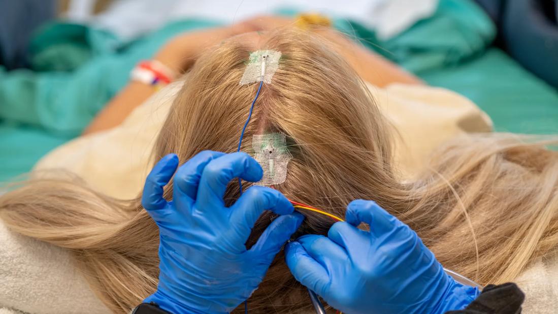
The case series consisted of 22 adults (8 women; mean age, 66.5 ± 11.2 years) treated for COVID-19 at Cleveland Clinic through May 20, 2020. Nineteen patients underwent cEEG with a 21-channel electrode system for at least 24 hours; three had routine EEG for less than one hour.
Six patients (27.3%) had a history of at least one neurologic comorbidity — i.e., epilepsy (2 patients), stroke (1), headache (1), traumatic brain injury (1) and spinal stenosis (2).
The hospital course involved the following:
EEG was ordered for altered mental status in 17 patients and for seizure-like events in five patients. These events consisted of left arm clonic movements, right face twitching (clonic), generalized tonic-clonic seizure and “eyebrow twitching with lip smacking.” Median duration of cEEG monitoring was 2 days (range, 1-6).
Advertisement
Among the 19 patients who underwent cEEG, findings were as follows:
Among the three patients who underwent routine EEG (none with a history of epilepsy), findings were as follows:
By the end of the study period, six patients (27.3%) had died during hospitalization and the rest had been discharged. No statistically significant associations were found between EEG findings and death.
Eighteen patients underwent brain CT and five underwent brain MRI. Except for a single case with acute symptomatic seizure, imaging findings did not correlate with epileptiform abnormalities or electrographic seizures on EEG.
Advertisement
The report in Neurocritical Care detailed two cases from the series, both of which involved men of advanced age with multiple comorbidities but no history of epilepsy who nevertheless experienced acute symptomatic seizures.
The first case was a 76-year-old who underwent cEEG for episodes of left upper extremity clonic activity and worsening encephalopathy. Monitoring revealed three focal seizures lasting about 30 seconds, each arising from the right centroparietal region. Clinical and EEG seizure activity subsided after levetiracetam initiation. He was ultimately discharged to a long-term acute care hospital.
The second case was an 82-year-old who underwent cEEG for right eyelid and facial twitching. Monitoring showed frequent EEG seizures from the left more than right frontal-temporal regions, which eventually progressed to status epilepticus. Most seizures were nonconvulsive. Seizure frequency improved with levetiracetam, but the patient died after being removed from life support 20 days into his ICU stay.
These reports reflect greater experience with COVID-19 patients showing evidence of seizures than has been reported elsewhere, Dr. Newey says, adding that this may be due to the long duration of cEEG conducted with a 21-channel electrode system.
The fact that EEG abnormalities correlated with neuroimaging findings in only one case suggests a direct link between COVID-19 and neurologic abnormalities, which Dr. Newey says is likely to involve multiple factors. “Seizures could indicate that COVID-19 is neurotropic or may possibly lead to microthrombi in the brain secondary to hypoxia or inflammation,” he conjectures. “It’s also possible that excessive cytokine activity disrupts the blood-brain barrier.”
Advertisement
Dr. Newey and his co-authors note that these reports also highlight the role of cEEG for evaluating COVID-19 patients with mental status changes and no explanatory neuroimaging findings. This utility may extend to helping guide therapy: Among the series’ six patients who had anti-seizure medications initiated during hospitalization, only two were discharged on them.
“Continuous EEG not only detected abnormal activity not seen by imaging but clarified which patients needed to continue anti-epileptic drugs,” Dr. Newey concludes. “This reduced the rate of unnecessary medication use.”
Advertisement
Advertisement
Patients report improved sense of smell and taste

Clinicians who are accustomed to uncertainty can do well by patients

Unique skin changes can occur after infection or vaccine

Cleveland Clinic analysis suggests that obtaining care for the virus might reveal a previously undiagnosed condition

As the pandemic evolves, rheumatologists must continue to be mindful of most vulnerable patients

Early results suggest positive outcomes from COVID-19 PrEP treatment
Could the virus have caused the condition or triggered previously undiagnosed disease?

Five categories of cutaneous abnormalities are associated with COVID-19