Large study gives guidance based on mental status, seizure etiology

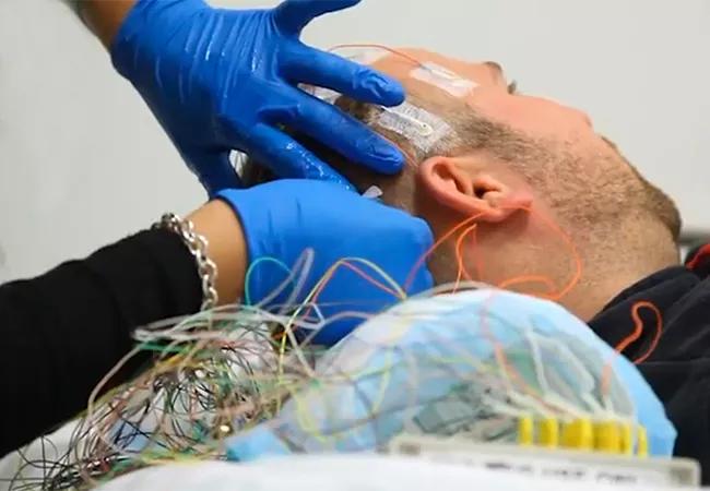
Among hospitalized patients requiring continuous EEG (cEEG) monitoring, those in a stuporous state and those with seizures due to hemorrhage, trauma or tumor are most likely to have delayed seizure detection and warrant longer cEEG monitoring. So concludes a large retrospective Cleveland Clinic study presented at the 2018 annual meeting of the American Academy of Neurology (AAN).
Advertisement
Cleveland Clinic is a non-profit academic medical center. Advertising on our site helps support our mission. We do not endorse non-Cleveland Clinic products or services. Policy
The findings represent a needed step toward better defining the optimal duration of cEEG monitoring according to patient characteristics and seizure etiology, explains the study’s senior author, Cleveland Clinic epileptologist Stephen Hantus, MD. “It’s now fairly widely recognized that there is a subpopulation of hospitalized patients who experience nonconvulsive seizures in the setting of another medical illness,” he says. “What we’re missing is the next step. How do you make smart choices about how long to monitor different patients to maximize detection of nonconvulsive seizures and actually improve care?”
Recognition of the prevalence of nonconvulsive seizures among hospitalized patients has emerged from several investigations in recent years, Dr. Hantus notes. One was an analysis by Cleveland Clinic’s cEEG monitoring group of 1,000 inpatients who underwent cEEG observation in 2009-2010; the aim was to determine who was at greatest risk for seizures. The researchers found that seizure detection rates increased in a linear fashion according to patient mental status — from 6 percent in patients with awake status to 20 percent with lethargic status to 25 percent with stupor to 33 percent with coma. The study also identified several high-risk etiologies for seizures: Stroke was most common, followed by hemorrhage, tumor, venous infection and approximately 25 less-common etiologies.
“This helped identify who should be monitored,” Dr. Hantus says. “But we have a limited number of monitoring machines, so we needed data-based guidance on how long to keep at-risk patients hooked up to a monitor.”
Advertisement
That’s where the current study comes in, which reviewed data for all inpatients at Cleveland Clinic’s main campus who underwent cEEG monitoring in 2016 (N = 2,425).
Among these 2,425 patients, 334 had seizures detected during monitoring. Median time to seizure onset was 3 hours, and 39 percent had their first seizure within an hour of monitoring. Nearly 14 percent had status epilepticus on cEEG.
For most seizure etiologies, at least 50 percent of patients had their seizures detected within the first 6 hours of cEEG monitoring. Seizures were detected within 24 hours among 80 percent of patients except those whose seizures were secondary to hemorrhage, trauma or tumor. Seizures were detected within 36 hours among 90 percent of patients except those whose seizures were due to hemorrhage, tumor or an unknown etiology.
Among all seizure etiologies, hemorrhage was the one most likely to be associated with seizure detection after 24 hours, particularly if a patient had more than one hemorrhage type.
When findings were evaluated according to patients’ mental status, seizures were detected within 2 days in 90 percent of patients who were awake, lethargic or comatose, whereas it took more than 3 days of monitoring for seizure detection in 90 percent of patients who were stuporous.
Overall, comatose patients and those with seizures secondary to cardiac arrest were most likely to have their seizures detected early.
“These findings suggest that if patients have one of these high-risk characteristics, such as stupor or hospitalization for hemorrhage (particularly more than one hemorrhage type), tumor or trauma, they merit longer cEEG monitoring to help ensure we’re not missing potential nonconvulsive seizures,” says Dr. Hantus.
Advertisement
He adds that more-intensive monitoring in these high-risk groups may also help flag patients at risk for epilepsy down the road, as about 50 percent of inpatients who have certain high-risk nonconvulsive seizure patterns such as periodic lateralized epileptiform discharges (PLEDs) during hospitalization will develop epilepsy after discharge. “We believe that longer cEEG monitoring in appropriate groups can allow us to improve long-term care and follow-up as well.”
In a separate study presented as a poster at the AAN meeting, Dr. Hantus and Cleveland Clinic colleagues took a deeper dive into one of the patient subtypes touched upon in the first study — hospitalized patients with intraparenchymal hemorrhage (IPH) who were monitored with cEEG.
“Among types of intraparenchymal hemorrhage, cortical hemorrhage is well recognized as a risk factor for nonconvulsive seizures, but the seizure risk posed by subcortical hemorrhages, which are far more common, is less clear,” Dr. Hantus notes.
To better define these risks, his team did a retrospective chart review of 121 patients with IPH who underwent brain CT or MRI as well as cEEG monitoring at Cleveland Clinic from January 2013 to December 2014. They found that a significant share of patients with IPH and seizures on cEEG had subcortical hemorrhages, accounting for 28 percent of cases with seizures and an abnormal EEG (defined as PLEDs).
“Subcortical intraparenchymal hemorrhage is a common form of IPH and often overlooked in terms of seizure risk, perhaps because most patients with subcortical IPH recover quite well,” says Dr. Hantus. “Yet these findings show that patients with supratentorial IPH, specifically subcortical IPH, should be evaluated for subclinical seizures, to help ensure that seizures get addressed properly if detected.”
Advertisement
Advertisement

Phase 2 trials investigate sitagliptin and methimazole as adjuvant therapies

Aim is for use with clinician oversight to make screening safer and more efficient

Rapid innovation is shaping the deep brain stimulation landscape

Study shows short-term behavioral training can yield objective and subjective gains

How we’re efficiently educating patients and care partners about treatment goals, logistics, risks and benefits

An expert’s take on evolving challenges, treatments and responsibilities through early adulthood

Comorbidities and medical complexity underlie far more deaths than SUDEP does

Novel Cleveland Clinic project is fueled by a $1 million NIH grant