Locations:

Shortcomings in specificity show influence of cortical changes beyond demyelination
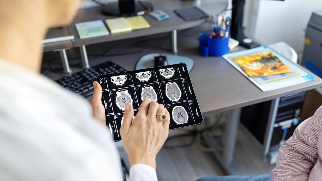
Several MRI modalities can be harnessed to better assess the severity and progression of multiple sclerosis (MS), but improved acquisition and post-processing methods are needed for reliable evaluation of cortical lesion load. So suggest new findings from Cleveland Clinic researchers published in Multiple Sclerosis Journal.
Advertisement
Cleveland Clinic is a non-profit academic medical center. Advertising on our site helps support our mission. We do not endorse non-Cleveland Clinic products or services. Policy
“Standard MRI methods are not specific enough to provide a complete picture of MS disease progression, especially for detecting damage within the cerebral cortex caused by cortical lesions,” says the study’s first author, Yufan Zheng, PhD, a postdoctoral fellow in the lab of Kunio Nakamura, PhD, in Cleveland Clinic’s Lerner Research Institute.
The myelin loss that is the hallmark of MS is particularly difficult to detect in the cerebral cortex, even with advanced MRI techniques. “We undertook this study to quantify how well these MRI techniques improve cortical lesion discovery,” Dr. Zheng explains.
The researchers examined how well three available advanced MRI modalities — MTR (magnetization transfer ratio), T1T2R (T1-weighted/T2-weighted ratio) and T2w (T2-weighted signal) — could determine areas of demyelination. They used these modalities to perform postmortem MRI studies on nine patients who had had primary or secondary progressive MS.
Following MRI acquisition, the researchers studied tissue samples from the cerebral cortices of each brain to determine how accurate the imaging techniques were in detecting cortical demyelination based on how much myelin proteolipid protein was present.
Of the three modalities, T2w was the most accurate in determining areas of demyelination (71% accuracy), followed by T1T2R (42%) and MTR (39%). T2w also had the highest specificity (46%) in detecting demyelination.
The effectiveness of T2w — particularly its role in determining T1T2R — can expand the effectiveness of MRI, Dr. Zheng notes. “We are excited about the possibility of T1T2R as a tool for mapping cortical myelin,” she says. “The fact that we can calculate the T1T2R map with routine T1w and T2w images makes T1T2R a possible new tool for myelin mapping.”
Advertisement
This has great potential for measuring the progression of MS, Dr. Zheng adds. “It’s important to be able to identify cortical lesions via MRI because they are now part of the MS diagnostic criteria,” she says.
While all three MRI techniques were relatively accurate in detecting areas of demyelination, their specificity was somewhat limited. Dr. Zheng believes this may be because conventional MRI is more sensitive to cortical changes other than demyelination. “This is why we need to improve our ability to use MRI in managing MS,” she says. “Higher-resolution imaging and better processing will go a long way in identifying changes in the cortex in these patients.”
The study was conducted by researchers in Cleveland Clinic’s Lerner Research Institute, Department of Neurosciences and Mellen Center for Multiple Sclerosis Treatment and Research. It was supported by the National Institute of Neurological Disorders and Stroke.
Advertisement
Advertisement

First full characterization of kidney microbiome unlocks potential to prevent kidney stones

Researchers identify potential path to retaining chemo sensitivity
Large-scale joint study links elevated TMAO blood levels and chronic kidney disease risk over time

Investigators are developing a deep learning model to predict health outcomes in ICUs.

Preclinical work promises large-scale data with minimal bias to inform development of clinical tests

Cleveland Clinic researchers pursue answers on basic science and clinical fronts

Study suggests sex-specific pathways show potential for sex-specific therapeutic approaches
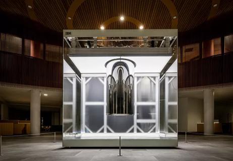
Cleveland Clinic launches Quantum Innovation Catalyzer Program to help start-up companies access advanced research technology