Locations:

Preclinical work promises large-scale data with minimal bias to inform development of clinical tests
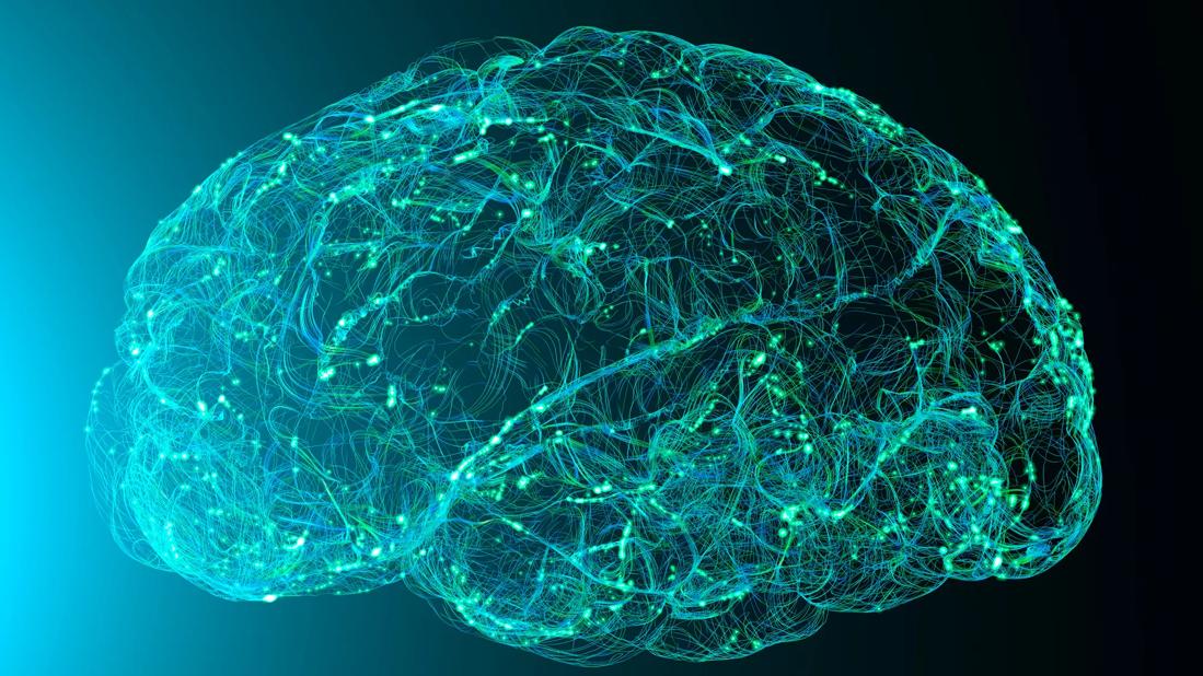
Image content: This image is available to view online.
View image online (https://assets.clevelandclinic.org/transform/632b71a6-64ec-4dff-801c-c0a2e5085c45/GettyImages-1219443537-jpg)
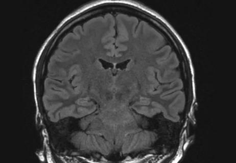
Brain mapping
A research team led by Cleveland Clinic and Oregon Health and Science University (OHSU) investigators has developed a new method for mapping the electrical paths that connect and coordinate various parts of the brain to complete behavioral and cognitive tasks. The findings promise insights critical to understanding behavior changes in patients with Alzheimer’s disease and other neurological disorders.
Advertisement
Cleveland Clinic is a non-profit academic medical center. Advertising on our site helps support our mission. We do not endorse non-Cleveland Clinic products or services. Policy
“Effects on behavior and personality in Alzheimer’s disease and related disorders are caused by changes in brain function,” says Hod Dana, PhD, of the Department of Neurosciences in Cleveland Clinic Lerner Research Institute, who is collaborating on the work with OHSU behavioral neuroscientist Jacob Raber, PhD. “If we can understand exactly how the changes occur, we may determine how to slow the process or stop it. Recording brain activity patterns that underlie behavioral changes is the first step to bridging the gap.”
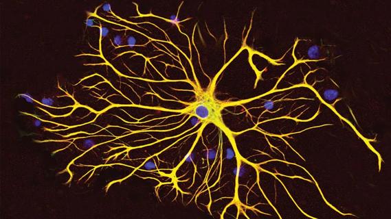
The team recently published results in Nature Communications (2023 Oct 12;14[1]:6399) on the use of a noninvasive calcium sensor system called CaMPARI (calcium-modulated photoactivatable ratiometric integrator) to map brain activity while a subject completes cognitive tasks. Drs. Dana and Raber are using CaMPARI in preclinical work to learn how genes related to Alzheimer’s disease affect the way neurons signal in the brain during learning and memory tasks.
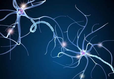
To study how brainwaves influence behavior and decision-making in mice, researchers observe as neurons turn “on” and “off” across the brain in different situations. However, current technologies for monitoring large-scale neuronal activity restrict animal movement and are unable to map the whole brain while still acquiring snapshots of activity at the single-cell level.
In contrast, CaMPARI-based recording can be done during free movement, thus capturing behavior, and can map single-cell activity. CaMPARI detects neuron activity by changing color from green to red inside active neurons during illumination with 400-nm light. After the test is completed, the red and green markers remain bright for several days, allowing the researchers to capture a series of images to track the brain’s activity by mapping where the red appears within the brain.
Advertisement
In the newly published study, the researchers used CaMPARI to record various cortical regions while a mouse completed a battery of behavioral and cognitive tests. They identified task-specific patterns of activity across motor and somatosensory cortices, observing significant differences among different parts of the motor cortex and correlations across certain activity patterns and tasks.
Dr. Dana says the free movement that CaMPARI enables is an advantage over current monitoring methods that fix a microscope to the animal’s head or require attachment of a recording device.
“We want to study the brain in its most natural state possible, without biasing it with our own tools,” he explains. “CaMPARI lets us map behavior-specific pathways in the brain without using obtrusive devices or limiting our field of view. It’s flexible and can be used to look at neuronal activity under various conditions. The results are reliable and reproducible, and it is easy to implement with commonly used scientific setups.”
Drs. Dana and Raber say their CaMPARI-based approach expands the capabilities of recording neuronal activity under minimally restricted conditions and provides large-scale behavioral data that can’t presently be generated otherwise. They hope to ultimately translate their results to develop tests and interventions that can improve quality of life for humans, providing patients with better treatment options.
“We now have the capability to study the relationship between brain activation and cognitive performance at an unprecedented level,” Dr. Raber notes. “These are the first steps in developing strategies to reverse changes and improve cognitive performance in people affected by neurological conditions.”
Advertisement
This work was funded by NIH R21AG065914 and U01NS123658.
Advertisement
Advertisement

Phase 3 STELLAR trial underscores role of molecular stratification in glioma care

A principal investigator of the landmark longitudinal study shares interesting observations to date
GFAP elevation may signal increased risk of progressive regional atrophy, cognitive decline
Research aims to extend observations of reversal learning in mice to human neurological disorders
Multicenter collaboration aims to facilitate tracking of neurological activity deep within tissue
Diagnosis and treatment of MOG antibody-associated disease

Designed for digitization, distance health, discovery and more
Model of viral encephalomyelitis shows links with CNS recovery from inflammation and damage