Locations:





Endovascular navigation technology clears first preclinical study
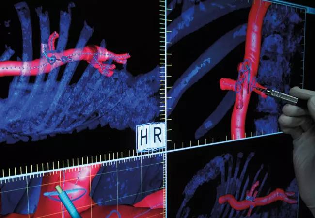
Image content: This image is available to view online.
View image online (https://assets.clevelandclinic.org/transform/7e67ed99-f224-4347-ba85-8d8d56e097db/16-HRT-1407-Centerline-IOPS-650x450_jpg)
Centerline Biomedical IOPS
Endovascular repair of aortic aneurysms and dissections is critically dependent on optimal understanding of the patient’s vasculature. To that end, Cleveland Clinic Innovations spinoff company Centerline Biomedical Inc. is refining development of its Intraoperative Positioning System (IOPS), a novel endovascular navigation technology developed by Cleveland Clinic researchers.
Advertisement
Cleveland Clinic is a non-profit academic medical center. Advertising on our site helps support our mission. We do not endorse non-Cleveland Clinic products or services. Policy
IOPS extracts the centerlines of the aorta and branch vessels from a patient’s CT and then mathematically constructs a high-quality three-dimensional model of the relevant vasculature, as shown in the image above. Electromagnetic tracking technology allows the model to be used in a GPS-like fashion to guide surgeons during minimally invasive endovascular aortic repairs, enabling less reliance on X-ray fluoroscopy.
The first preclinical in vivo study of IOPS was completed in May 2016 at Cleveland Clinic Lerner Research Institute. It demonstrated the system’s ability to provide non-radiation-based navigation with superior visualization compared with X-ray fluoroscopy, which is limited by its two-dimensional visualization and by the radiation exposure it confers on patients and providers.
“Preclinical evaluation verified the ability to use this navigation system in manipulating through the aorta and its branches in the absence of radiation-inducing fluoroscopy,” says Cleveland Clinic vascular surgeon Matthew Eagleton, MD, who led the study. Noting that catheters and guidewires were easily visualized on the IOPS display, he predicted that the system “will be a game changer in the era of endovascular therapy.”
Centerline Biomedical plans to submit IOPS for FDA review, with a target market entry in the third quarter of 2017.
Advertisement
Advertisement

AHA statement details nuanced, evidence-based strategies
The benefits of multiarterial grafting are possible without opening the chest
Collaborative citywide initiative reshapes protocols to boost cardiac arrest survival
For severe cases, complete unroofing of the bridge is imperative
Lower threshold expanded revascularization candidacy without raising early complication rates

Elevated levels of the gut microbial metabolite predict prevalence and adverse outcomes in community-based study
They generally depend on the mutated gene, clone size and clinical setting
Large analysis adds to evidence that absolute diameter alone may miss vulnerable patients
 Search