Locations:

How it adds value in structural assessment of bioprosthetic MV function
By Paul Cremer, MD
Advertisement
Cleveland Clinic is a non-profit academic medical center. Advertising on our site helps support our mission. We do not endorse non-Cleveland Clinic products or services. Policy
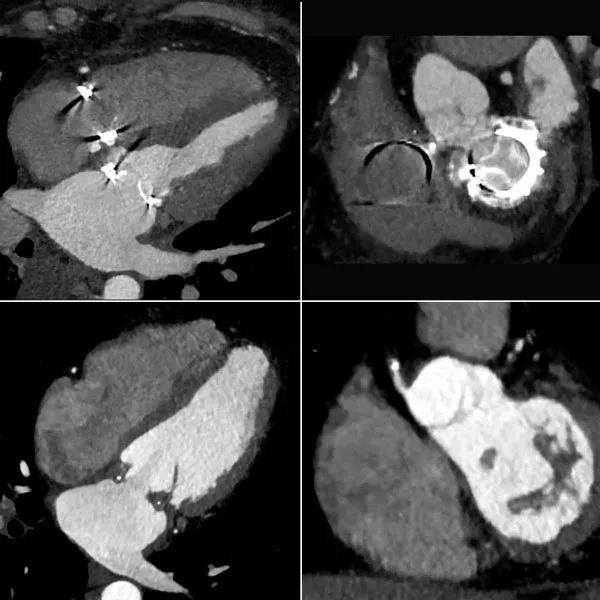
Image content: This image is available to view online.
View image online (https://assets.clevelandclinic.org/transform/9a1ab1a4-842b-453a-b573-c09adcd62985/17-HRT-3918-Cremer-inset_jpg)
Echocardiography is the mainstay for assessing bioprosthetic mitral valve function, but when it comes to assessing structure, there’s an emerging role for four-dimensional (4-D) CT. That’s particularly the case when there’s a diagnostic question of whether the predominant pathology is calcification versus valve thrombosis.
The 4-D CT images above highlight this point. The top two images are from a patient with symptomatic bioprosthetic mitral valve stenosis. On the left, the reconstructed apical four-chamber view shows hyperattenuated bioprosthetic valve leaflets, representing severe calcification. On the right, the short-axis reconstruction also shows leaflet hyperattenuation, consistent with calcification.
In contrast, the bottom two images are from a case of subacute bioprosthetic valve thrombosis in a patient with symptomatic mitral stenosis. On the left, the apical four-chamber reconstruction shows layering hypoattenuation along the sewing ring, indicating thrombus. On the right, the short-axis reconstruction shows prominent hypoattenuation and increased leaflet thickness illustrating the extension of thrombus onto the leaflets.
Imaging calcium is a strength of CT and a relative weakness of echocardiography, despite the overall strength of echo to assess the severity of bioprosthetic mitral valve dysfunction. The above images highlight how helpful 4-D CT can be in evaluating structural causes of bioprosthetic mitral valve dysfunction, particularly for distinguishing between calcification and thrombus.
Advertisement
Dr. Cremer (cremerp@ccf.org) is a cardiologist in Cleveland Clinic’s Section of Cardiovascular Imaging in the Sydell and Arnold Miller Family Heart, Vascular and Thoracic Institute.
For more reading on how 4-D imaging informs complex aortic valve repair in adult and pediatric patients, read this story from Cleveland Clinic Children’s of how a congenital heart surgeon and heart anatomist-imager team up to improve surgical success.
Advertisement
Advertisement

New framework better distinguishes stable from critically ill patients

CMR-CLIP outperforms general AI tools; may one day expand patient access to CMR

Post hoc analysis of CLEAR Outcomes trial bolsters its case as a statin alternative

Large retrospective analysis may prompt prospective studies

How to talk about lifetime risk, treatment goals, Lp(a) testing, statin skepticism and more

A scannable recap of recent volumes and clinical metrics from Cleveland Clinic

Cleveland Clinic reports first U.S. series focused on use in this challenging setting

Large series confirms early and long-term survival advantages over partial pericardial resection