Locations:

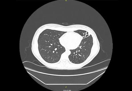
Not all lung nodules are malignant
Not all lung nodules are malignant. In this video, Sanjay Mukhopadhyay, MD, staff pathologist in the Department of Anatomic Pathology, compares and contrasts two cases of lung granulomas caused by fungal infection. These types of fungal infection cause granulomas in the lung, which can be mistaken for each other.
Advertisement
Cleveland Clinic is a non-profit academic medical center. Advertising on our site helps support our mission. We do not endorse non-Cleveland Clinic products or services. Policy
Video content: This video is available to watch online.
View video online (https://www.youtube.com/embed/aRuH_GwPYyA?feature=oembed)
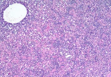
He starts the first case with whole slide imaging of a lung nodule performed to rule out malignancy. At low magnification, the lung parenchyma appears normal with a visible interlobular septum. The periphery of the nodule is cellular, and the center of the nodule appears necrotic even at low magnification. At higher magnifications, the necrosis is the type that one might expect to see in tuberculosis, non-tuberculous mycobacterial infection or fungal infection. The periphery of the lesion appears granulomatous as the cells are actually epithelioid histiocytes tightly clustered together with a few multinucleated giant cells. The lesion is then a necrotizing granuloma, and differential diagnosis first and foremost includes mycobacterial or fungal infection. Mycobacteria will not be visible on hematoxylin and eosin (H&E) stains, but fungi may be. The four most common fungi found in necrotizing granulomas are Histoplasma, Cryptococcus, Blastomyces and Coccidioides. The last three can be seen on H&E stains.
Dr. Mukhopadhyay examines the necrosis but cannot find any fungi at the highest possible magnification. He then examines Grocott’s methenamine silver (GMS) stain. He magnifies the necrotic area and points out the numerous Histoplasma organisms stained black within the necrosis. Watch the video to see the second case.
Cleveland Clinic Laboratories’ Pathology Insights video series features important cases, methods, and practices that are personally presented by our staff pathologists.
Advertisement
These short videos break down information about interesting pathology cases to better inform doctors, laboratory staff, patients or anyone interested in the field of pathology.
Advertisement
Advertisement

Screen patients seeking care for chlamydia, gonorrhea
Lingulectomy removes infection when antibiotics fail

Researchers have developed immunoprofiles for an emerging disease with a mortality rate as high as 27%

Findings from one of the first published case series
Don't discount this crucial step
EBUS-TBNA found safe and effective
How to spot the rare infection

An uncommon condition in a 41-year-old man