Research to investigate non-invasive method aims to identify patients at risk for osteoarthritis

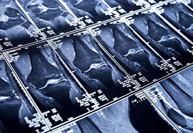
Xiaojuan Li, PhD, director of the Program of Advanced Musculoskeletal Imaging (PAMI), has received a five-year, $3 million grant from the National Institute of Arthritis and Musculoskeletal and Skin Diseases to develop a quicker method to assess changes in the knee that lead to osteoarthritis (OA).
Advertisement
Cleveland Clinic is a non-profit academic medical center. Advertising on our site helps support our mission. We do not endorse non-Cleveland Clinic products or services. Policy
Her new, quantitative magnetic resonance imaging (MRI) methods — a refinement and improvement over traditional MRI techniques — are slated to more quickly and efficiently classify at-risk patients, which may allow physicians to intervene earlier with behavior modification or other treatments to prevent or delay OA symptoms.
OA is a common and sometimes debilitating disease that affects more than 27 million people in the United States and often manifests in the knee. Clinical imaging evaluation of OA has relied primarily on plain radiography, which only depicts bone-related changes that occur late in the disease. Unlike plain radiography, MRI is a powerful modality for evaluating soft tissues such as cartilage and menisci.
While significant efforts have been made in the past decade to develop quantitative MRI to detect early tissue degeneration in OA, existing methods are time-intensive and costly, and the equipment’s capabilities often vary with different manufacturers.
With this new award, Dr. Li and her team will conduct studies in patients with anterior cruciate ligament (ACL) tears, a common indicator of knee OA. She will develop a novel process to quantify biological markers of cartilage degeneration and test the method across multiple protocols and vendors to demonstrate its utility in the clinical setting.
To bypass the traditional limitations of MRI in diagnosing OA, Dr. Li and her team are building on the premise that specific cartilage measurements (called MR T1p and T2 measurements) provide a more sophisticated and sensitive method to assess biochemical changes within the cartilage matrix, and may be promising biomarkers for early cartilage degeneration. Dr. Li’s team plans to develop new, accelerated T1ρ and T2 imaging methods and will test them on MRI systems from three manufacturers at four sites.
Advertisement
“There are currently no disease-modifying treatments for OA, so it is critical to be able to identify markers of disease for early intervention,” says Dr. Li. “We hope that our method of advanced, fast quantitative cartilage imaging will help transform OA management in patients.”
The ultimate goal, she says, will be to create a standard for MRI usage in future clinical trials of patients who have OA. When the study is complete, Dr. Li hopes to have in place an improved method of detecting early joint degeneration and predicting future OA.
Dr. Li was recruited to Cleveland Clinic in 2017, where she is staff in the Department of Biomedical Engineering and holds the Bonutti Family Endowed Chair for Musculoskeletal Research.
This article was originally featured on the Lerner Research Institute website.
Advertisement
Advertisement

First full characterization of kidney microbiome unlocks potential to prevent kidney stones

Researchers identify potential path to retaining chemo sensitivity

Large-scale joint study links elevated TMAO blood levels and chronic kidney disease risk over time

Investigators are developing a deep learning model to predict health outcomes in ICUs.

Preclinical work promises large-scale data with minimal bias to inform development of clinical tests

Cleveland Clinic researchers pursue answers on basic science and clinical fronts

Study suggests sex-specific pathways show potential for sex-specific therapeutic approaches

Cleveland Clinic launches Quantum Innovation Catalyzer Program to help start-up companies access advanced research technology