Locations:

DT-MRI and RNA-FISH show increase in CITED4 promotes healthy architecture of myocardium

Enhancement of the gene transactivator CITED4, stimulated by exercise, is key to strengthening myocardium by promoting a complex helical structural pattern of myocytes. So found a series of investigations in mice published in Communications Biology (2022;5[1]:656) using diffusion-tensor magnetic resonance imaging (DT-MRI) and RNA fluorescence in situ hybridization (RNA-FISH). Elucidating this pathway in humans may one day lead to more personalized exercise “prescriptions” and other interventions that mimic the effects of exercise.
Advertisement
Cleveland Clinic is a non-profit academic medical center. Advertising on our site helps support our mission. We do not endorse non-Cleveland Clinic products or services. Policy
“These are the first studies to identify microstructural changes occurring on a tissue level as a result of specific regional gene expression,” says Christopher Nguyen, PhD, Director of MRI Research at Cleveland Clinic and a corresponding author of the study. “We have known for a long time that exercise is good for the heart, but these studies may indicate why.”
The therapeutic benefit of exercise to the heart is not well understood on a cellular level. Previous studies have shown that exercise induces changes in myocytes, increasing their length and width. This beneficial hypertrophy is distinct from the better known pathological cardiac hypertrophy, in which myocyte length increases disproportionately.
The effects of these differences on structural remodeling of myocardium are difficult to study. Laborious histological sectioning led to the discovery that myocardium has a helical microstructure, with orientation of fibers smoothly transitioning from right-handedness to left-handedness through the tissue layers between the endocardium and epicardium. This double helical pattern confers maximal strength and is associated with improved outcomes following cardiac injury and disease.
Using 14T DT-MRI and techniques that Dr. Nguyen’s team developed, the investigators can more easily and noninvasively map the underlying orientation of cardiomyocytes and thereby detect structural changes (see recent Consult QD post on cardiac MRI innovations) while keeping the tissue intact. This study also made use of RNA-FISH to study the spatial distribution of the RNA transcript of CITED4, a protein that increases the rate of gene expression and has been implicated in exercise-induced cell hypertrophy pathways.
Advertisement
The two novel technologies — DT-MRI, which provides high-resolution images of tissue architecture, and RNA-FISH, which allows quantification and localization of gene expression (i.e., RNA and protein molecules) within tissue — have enabled the current study investigators to assess the impact of exercise on heart muscle microstructure in detail for the first time. Both wild-type and genetically modified floxed CITED4 mice were used to reveal these fundamental findings.
The investigations involved mice subjected either to exercise (free access to a running wheel) or to sedentary conditions for eight weeks, as follows:
After eight weeks, hearts were extracted, and exercised mice were found to have greater myocardial wall thickness and left ventricular mass compared with sedentary mice. Spatial distribution and quantity of CITED4 were analyzed using RNA-FISH and compared with tissue microstructure using DT-MRI.
Among the findings:
Advertisement
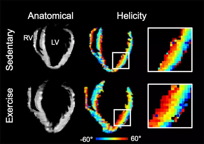
Image content: This image is available to view online.
View image online (https://assets.clevelandclinic.org/transform/d53ec01a-2dd9-44e9-b7d7-f49ac4412d43/22-HVI-3073821-CQD-inset_jpg)
Figure. Representative anatomical images and helicity maps of sedentary and exercise subjects revealing how the heart enlarges and the preferential increase in helicity in the lateral wall. Note the increased dark red region representing an increase in right-handed helical heart fibers in the zoomed inset.
Dr. Nguyen highlights the following key points of these investigations:
“New genetic and imaging technologies can be powerfully combined to help us understand how the body — and medical interventions — work at a cellular level,” Dr. Nguyen notes. “Furthermore, because MRI is a noninvasive technology being used in the clinic, we can more rapidly start human trials following initial animal studies.”
Dr. Nguyen says his team is planning future studies in patients with cardiovascular disease to characterize changes in cardiac architecture after an exercise program.
Advertisement
“The associations of exercise, CITED4 upregulation and cardiac microstructure benefits may have important clinical implications,” says Dr. Nguyen. “It is possible that one day we will be able to target delivery of CITED4 or other modifiers of cardiac tissue to specific areas of the heart. In the meantime, this is one more piece of evidence of the power of exercise for a healthy heart.”
“While multiple studies have demonstrated the significant benefits of exercise and cardiac rehabilitation programs for patients with significant cardiac disease, this fascinating study elucidates the underlying mechanism and the ability to visualize why the heart gets stronger with exercise,” adds Deborah Kwon, MD, Director of Cardiac MRI at Cleveland Clinic.
Advertisement
Advertisement

First full characterization of kidney microbiome unlocks potential to prevent kidney stones

Researchers identify potential path to retaining chemo sensitivity
Large-scale joint study links elevated TMAO blood levels and chronic kidney disease risk over time

Investigators are developing a deep learning model to predict health outcomes in ICUs.

Preclinical work promises large-scale data with minimal bias to inform development of clinical tests

Cleveland Clinic researchers pursue answers on basic science and clinical fronts

Study suggests sex-specific pathways show potential for sex-specific therapeutic approaches
Cleveland Clinic launches Quantum Innovation Catalyzer Program to help start-up companies access advanced research technology