Locations:

Noninvasive, radiation-free imaging supports treat-to-target IBD care
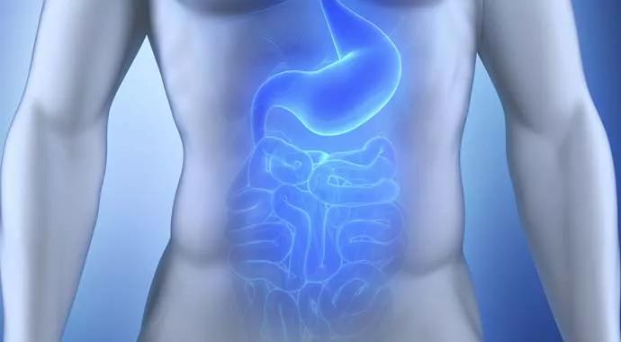
Image content: This image is available to view online.
View image online (https://assets.clevelandclinic.org/transform/6ca4aa59-568a-45fa-a063-514f6fea4269/Gastroparesis-Clinic-690x380-jpg)
Noninvasive, radiation-free imaging supports treat-to-target IBD care
Intestinal ultrasound (IUS) has emerged as a noninvasive, radiation-free alternative for tracking disease activity that is helping clinicians provide treat-to-target care for patients with inflammatory bowel disease (IBD).
Advertisement
Cleveland Clinic is a non-profit academic medical center. Advertising on our site helps support our mission. We do not endorse non-Cleveland Clinic products or services. Policy
It is estimated that between 2.4 and 3.1 million people in the United States have IBD, a chronic, relapsing-remitting inflammatory condition for which there is no cure. The most common forms of IBD are Crohn's disease and ulcerative colitis, both of which require continuous monitoring to assess disease activity and guide treatment decisions.
According to a 2024 report from the American Gastroenterological Association (AGA), a growing number of providers are incorporating IUS for the management of IBD. That includes board-certified gastroenterologist Asad ur Rahman, MD, Medical Director of the Inflammatory Bowel Disease Center at Cleveland Clinic in Florida, one of the few IBD referral centers in southeast Florida.
“Intestinal ultrasound provides a detailed, real-time view of the bowel that supports an enhanced level of monitoring while reducing exposure to the risks and burdens associated with traditional imaging techniques,” states Dr. Rahman. He is the first within Cleveland Clinic’s five-hospital regional health system in Florida to use point-of-care IUS as part of a broader monitoring strategy for IBD.
IUS requires two different ultrasound probes to capture both global and detailed views of the bowel. The first is a low-frequency curvilinear probe that provides a general view of the entire bowel and helps identify areas of significant concern. A high-frequency linear probe is then used to zoom in on specific segments of the intestine and measure critical indicators of disease activity.
Advertisement
“We look at a number of factors, including bowel wall thickness and blood flow to the bowel wall, both of which are key signs of active inflammation,” explains Dr. Rahman. He notes bowel wall thickness, in particular, is a sensitive indicator of active inflammation. A normal adult bowel wall measures 3 mm or less, so anything over 3 mm is considered abnormal.
IUS is also used to assess stratification of the five bowel wall layers, the presence of strictures, fistulas, and abscesses, and mesenteric fat proliferation, which can accumulate around the bowel wall in the presence of inflammation.
“Advancements in ultrasound technology have improved the resolution of the images captured and our ability to see disease activity, making it very useful for monitoring patients with IBD,” says Dr. Rahman. “IUS typically takes less than 30 minutes to perform, which also makes it a convenient point-of-care option that is noninvasive and repeatable.”
While not a standalone diagnostic tool for IBD – since endoscopic and histologic confirmation are still required – IUS enhances clinical decision-making by providing objective data on disease extent and severity. “It is particularly useful for examining the ileum and colon, with the exception of the rectum, and for establishing a baseline of disease activity,” says Dr. Rahman.
One of the most significant advantages of IUS is its ability to track disease activity over time through quantifiable markers such as bowel wall thickness and vascularity. These indicators closely correlate with inflammation and allow clinicians to detect subtle disease changes and track disease progression beyond symptom reporting.
Advertisement
Over the past decade, IBD management has shifted away from clinical symptom management alone toward objective disease measurement and treat-to-target strategies, as recommended by the Selecting Therapeutic Targets in Inflammatory Bowel Disease (STRIDE) guidelines published in 2015. Authors of the subsequent STRIDE-II update in 2021 also recognized bedside IUS as a meaningful tool for repeated assessment of treatment response, further integrating it into routine clinical practice.
“IUS supports early detection of disease flare-ups or complications and enables treatment decisions based on the most current and precise data available,” says Dr. Rahman. “It also offers a fast, accessible way to guide therapy adjustments without relying solely on symptoms or invasive procedures.”
Comparative data cited in the AGA’s most recent clinical practice update indicate that IUS matches the accuracy of computed tomography enterography (CTE) and magnetic resonance enterography (MRE) in monitoring IBD and identifying disease complications. Studies also have shown that IUS has superior predictive capabilities than other imaging modalities and the ability to assess a deeper degree of remission observed through transmural healing.
In clinical practice, Dr. Rahman says IUS has proven particularly valuable when other tools fall short. In one case, a young woman with abdominal pain had a normal colonoscopy and a nonspecific CT finding. IUS revealed transmural thickening and fat wrapping in the terminal ileum, prompting further workup and a diagnosis of Crohn’s ileitis. After initiating therapy, the patient saw significant improvement, allowing her to return to college.
Advertisement
“I think intestinal ultrasound really added value to her care,” Dr. Rahman reflects. “It validated what she was experiencing and led to a diagnosis.”
Clinically, IUS is quick, accessible, and does not require any special preparation. This allows for point-of-care assessments during routine visits, enabling timely evaluation of disease activity without the delays or logistical complexity of more invasive tests. “Patients do not undergo bowel prep or sedation, which improves comfort and adherence to follow-up protocols,” says Dr. Rahman.
Unlike traditional imaging techniques, IUS does not require radiation or contrast agents, making it a safer option, especially for younger patients and those needing frequent monitoring. It also reduces the cumulative risk associated with repeated imaging and contributes to lower overall healthcare costs.
Importantly, IUS provides real-time visualization of disease activity. This immediacy allows clinicians to assess therapeutic response on the spot and supports dynamic treatment decisions.
From a patient engagement perspective, IUS also offers a unique opportunity for patients to view live images of their intestines. Studies suggest this can help patients better understand their condition.
“This interactive approach fosters shared decision-making, which I think is a really important benefit,” adds Dr. Rahman. “I’ve found that when my patients are more involved in their treatment decisions, their compliance tends to improve.”
Taken together, the safety, speed, accessibility, and patient-centered nature of IUS make it an increasingly indispensable tool in modern IBD care, confirms Dr. Rahman.
Advertisement
Because IUS cannot distinguish between IBD and other causes of colonic or small bowel inflammation, such as infectious or drug-induced colitis, colonoscopy remains the gold standard for definitive diagnosis and tissue sampling.
Dr. Rahman points out that direct visualization of the mucosal surface and targeted biopsy are essential for dysplasia surveillance. It is the primary tool used to monitor disease activity for people who have a risk of dysplasia or a previous history of dysplasia.
“Patients with primary sclerosing cholangitis as well as other patients with longstanding inflammation should undergo colonoscopy with biopsies to assess for dysplasia,” he says. “For patients who have not had longstanding inflammation and have achieved endoscopic healing, IUS is a good test for ongoing monitoring.”
Dr. Rahman expects that IUS will become more widely adopted over the next several years, expanding beyond the academic health systems, like Cleveland Clinic, that have embraced the technology in the past three to four years.
He points to the efforts of the International Bowel Ultrasound Group (IBUS) and the Intestinal Ultrasound Group of the United States and Canada (iUSCAN). Both nonprofits are focused on IUS education, training, research and advocacy in IBD care.
“I think IUS will become a mainstay of monitoring disease activity and will probably replace the need for a lot of cross-sectional CT and MRI scans,” comments Dr. Rahman. “We'll also likely be able to avoid annual colonoscopies for many patients, and limit its use in ongoing care to dysplasia surveillance and the evaluation of precancerous changes.”
As Associate Program Director of the Gastroenterology Fellowship at Cleveland Clinic in Florida, Dr. Rahman is also committed to educating the next generation of gastroenterologists on its benefits. He believes IUS will be an important addition to the fellowship curriculum.
“Intestinal ultrasound does not completely replace established imaging techniques. The key to maximizing its benefit lies in integrating IUS appropriately into the broader IBD monitoring strategy, taking into account each patient’s unique needs and risk factors,” he concludes.
Advertisement

Study highlights strong predictive value of circulating tumor DNA testing

Researchers with Cleveland Clinic in Florida highlight need for a national registry for gastrointestinal stromal tumors

Multidisciplinary LARS Center addresses life-altering consequence of rectal cancer surgery

Early recognition and intervention recommended in cubital tunnel syndrome

Manpreet (Meena) Bedi, MD, named Division Chair of Radiation Oncology

AR-assisted navigation is closing the gap between surgical planning and implant placement

Evidence shows early evaluation improves survival and quality of life – yet many eligible patients are referred too late

2026 ADA Standards of Care promote holistic, multisystem management