Locations:

Marasco lab brings sensation to the fore in prosthetics development
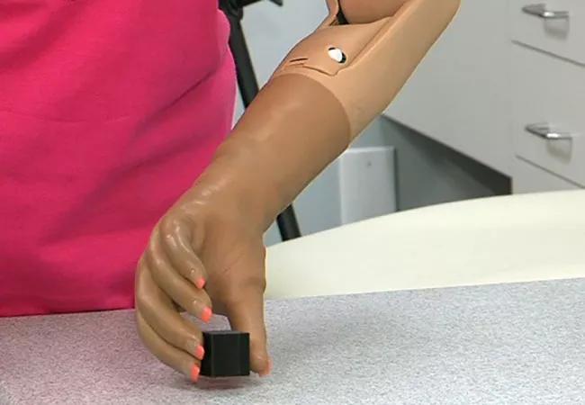
Image content: This image is available to view online.
View image online (https://assets.clevelandclinic.org/transform/bb2f00c9-1677-4dd8-b83f-1b151c50c06a/17-NEU-654-Marasco-650x450_jpg)
17-NEU-654-Marasco-650×450
Paul Marasco, PhD, is a researcher who seems to have the touch these days. Not only was he a recipient of the 2016 Presidential Early Career Award for Scientists and Engineers — the highest honor bestowed by the U.S. government on early-career professionals in these disciplines — but he’s in the vanguard of research efforts in advanced upper limb prosthetics, as detailed in this earlier post.
Advertisement
Cleveland Clinic is a non-profit academic medical center. Advertising on our site helps support our mission. We do not endorse non-Cleveland Clinic products or services. Policy
Moreover, touch is at the heart of Dr. Marasco’s contributions to his field, as the presidential award recognized his work studying use of neural-machine interfaces to provide touch and movement sensations to prosthetic limbs.
“We’re trying to integrate touch so that amputees feel as if their prosthetic limbs are part of their body,” explains Dr. Marasco, who pursues that work in his dual roles as principal investigator for the Advanced Platform Technology Center of Excellence at the Louis Stokes Cleveland Veterans Affairs Medical Center and as associate staff in Cleveland Clinic’s Department of Biomedical Engineering.
Specifically, his team provides amputee research subjects with artificial limbs fitted with sensors and tiny electronic robots that transfer impulses from the artificial hand or arm to a site with small motors on the chest or upper arm where the nerves that once led to the lost limb have been surgically relocated and reinnervated. The new nerve site is mapped according to various movement, position and touch sensations. This connects stimuli from the limb to the appropriate relocated nerve leading to the brain, thereby communicating limb movement and touch sensation feedback to the patient.
“We’ve found that the brain involuntarily incorporates the prosthetic limb into the subject’s self-image,” Dr. Marasco says. “No training is needed.”
“Sensation means everything,” adds Frederick Frost, MD. “Decades of medical research have shown that we can make prosthetics — and even paralyzed limbs — move with tendon transfers and electricity. But patients won’t use the technology if they have to depend on their eyes to see what their hand is doing.”
Advertisement
Dr. Marasco’s team has used this approach to explore sensory feedback in seven reinnervated research participants to date across the U.S. and Canada. For one study participant, Claudia Mitchell, “the sensation piece has been huge,” as she explains in the two-minute video below.
Video content: This video is available to watch online.
View video online (https://www.youtube.com/embed/IrXiXcefRO0?feature=oembed)
Dr. Marasco’s research team will be directing future efforts toward applying this cognitive- and perceptual-based technology to individuals with loss of sensation due to conditions such as diabetes and spinal cord injury. “An additional focus will be how sensation interacts with motor control, with an eye toward maximizing sensation in relation to movement,” he says.
He attributes much of his team’s progress to their distinctive opportunity to work as both a science lab and a clinical lab, as he explains in the one-minute video below.
Video content: This video is available to watch online.
View video online (https://www.youtube.com/embed/0Fk4P0bTjVU?feature=oembed)
Advertisement
Advertisement

Findings may have implications for understanding the disorders’ pathophysiology

Routine capture of standardized neuroperformance data may expand and refine investigations

Nearly one-fifth of such cases fulfill 2024 McDonald criteria based on biomarkers

Adequate dosing may improve outcomes in well-selected patients, large Cleveland Clinic series suggests

Reimagining the outpatient neurological visit with routine capture of neuroperformance data

Free portal helps researchers classify and share data using the IC-CoDE framework

Large study shows rural patients are less apt to be discharged to inpatient rehab, hampering outcomes

Updates on this fast-evolving therapeutic landscape from a leading trialist