Diffuse alveolar damage in acute respiratory distress syndrome
Video content: This video is available to watch online.
View video online (https://www.youtube.com/embed/3usFHIHCr2c?feature=oembed)
Diffuse Alveolar Damage in ARDS
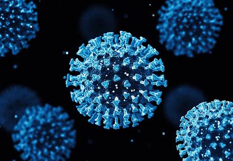
For most patients who are infected with the SARS-CoV-2 virus (the disease caused by this virus is referred to as COVID-19), symptoms are typically mild-to-moderate, often restricted to the upper respiratory system. However, in more severe cases, patients can develop acute respiratory distress syndrome (ARDS), leading to a worse prognosis.
Advertisement
Cleveland Clinic is a non-profit academic medical center. Advertising on our site helps support our mission. We do not endorse non-Cleveland Clinic products or services. Policy
The onset of ARDS, a noncardiogenic pulmonary edema, begins with viral entry into alveolar pneumocytes, which line the alveoli, the gas exchange units of the lungs. These cells are also vulnerable to infection from influenza. The alveolus resembles a balloon-like structure (featured in the video), which facilitates gas exchange in the lungs. However, once the virus permeates this sac, the cells will eventually experience apoptosis, triggering cell death in that alveolar unit, as well as adjacent units. This process leads to diffuse alveolar damage (DAD), injuring the capillaries that deliver oxygen to the rest of the body, as well as other cells in the alveoli.
The damage to capillaries and other alveolar cells in DAD combines to produce a form of debris known as hyaline membranes, a key pathologic feature of the disease. The thickening of the alveolar wall impedes diffusion of oxygen into the capillaries, making it very difficult for patients to breathe. Patients with DAD also experience acute edema and inflammation. This process is thought to be the underlying basis of the development of ARDS in patients with COVID-19.
In addition to disruptions in alveolar-capillary permeability, ARDS is characterized by reduced alveolar clearance and collapse, increased pulmonary vascular resistance, reduced compliance, the development of edema, and gas exchange abnormalities. It can necessitate extra corporeal membrane oxygenation or transplant. It can also cause cerebrovascular injury, which may be more common in the setting of COVID-19 according to a 2022 comparison of brain MRI findings led by Cleveland Clinic physicians.
Advertisement
Advertisement

Patients report improved sense of smell and taste

Clinicians who are accustomed to uncertainty can do well by patients

Unique skin changes can occur after infection or vaccine

Cleveland Clinic analysis suggests that obtaining care for the virus might reveal a previously undiagnosed condition

As the pandemic evolves, rheumatologists must continue to be mindful of most vulnerable patients

Early results suggest positive outcomes from COVID-19 PrEP treatment

Could the virus have caused the condition or triggered previously undiagnosed disease?

Five categories of cutaneous abnormalities are associated with COVID-19