Locations:

A pathologist diagnoses a recently described form of IF
In this video, Sanjay Mukhopadhyay, MD, staff pulmonary pathologist in the Department of Anatomic Pathology, presents a case of a recently identified form of interstitial fibrosis known as Smoking-Related Interstitial Fibrosis (SRIF). He also explains respiratory bronchiolitis, an indication that the patient is or was a smoker, and how its identification from pigment-filled macrophages can assist in diagnosis.
Advertisement
Cleveland Clinic is a non-profit academic medical center. Advertising on our site helps support our mission. We do not endorse non-Cleveland Clinic products or services. Policy
Video content: This video is available to watch online.
View video online (https://www.youtube.com/embed/_7MDaZ-mces?feature=oembed)
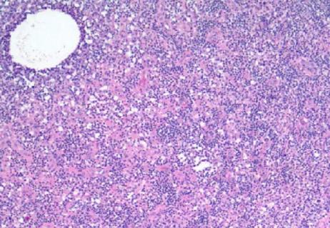
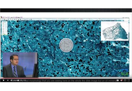
Dr. Mukhopadhyay shows an image from a transbronchial biopsy and identifies alveolated lung in the background. He also identifies the alveolar septum and pigmented macrophages within the air spaces. At higher magnification, these macrophages are light brown and occasionally have dark black specks. These Dr. Mukhopadhyay identifies as an important clue leading to a diagnosis of respiratory bronchiolitis.
He explains that respiratory bronchiolitis is a confusing term because it does not mean “inflammation of the bronchioles,” but it does not necessarily need to involve bronchioles and the cell of interest is macrophages rather than lymphocytes or plasma cells. Respiratory bronchiolitis is rather defined by the accumulation of pigment-filled macrophages within any air spaces within the lumens of respiratory bronchioles, alveolar ducts or alveoli. The pigment comes from cigarette smoke, and the presence of bronchiolitis indicates the patient is or was a smoker. Nine years can pass between ceasing smoking and lung biopsy, and respiratory bronchiolitis can still be present.
Dr. Mukhopadhyay notes that it is important to distinguish the brown pigment characteristic of respiratory bronchiolitis from hemosiderin. The characteristic look of respiratory bronchiolitis is finely granular brown pigment along with an occasional black speck at high magnification. The particles for hemosiderin are a bit coarser and larger with no black specks. He shows the two side by side to highlight the difference and notes that iron staining is not a foolproof way to distinguish between the two. Watch the video for more pearls of wisdom from Dr. Mukhopadhyay.
Advertisement
Advertisement

Screen patients seeking care for chlamydia, gonorrhea
Lingulectomy removes infection when antibiotics fail

Researchers have developed immunoprofiles for an emerging disease with a mortality rate as high as 27%

Findings from one of the first published case series
Don't discount this crucial step
EBUS-TBNA found safe and effective
How to spot the rare infection
Not all lung nodules are malignant