Locations:

Researchers pair postmortem MRI with histopathology to explore thalamic atrophy
In patients with multiple sclerosis (MS), degeneration of thalamic projections and/or a neurodegenerative process appears to make a greater contribution to thalamic atrophy than thalamic demyelinating lesions, according to Kedar Mahajan, MD, PhD, a neurologist in Cleveland Clinic’s Department of Neurosciences and Mellen Center for MS Treatment and Research.
Advertisement
Cleveland Clinic is a non-profit academic medical center. Advertising on our site helps support our mission. We do not endorse non-Cleveland Clinic products or services. Policy
He and Cleveland Clinic colleagues conducted postmortem in situ MRI studies in 95 subjects with MS and compared thalamic lesion volume with pathologic examination findings identifying thalamic demyelination and microglial/macrophage activation. Results were published in the Annals of Neurology (2020 Apr 14 [Epub ahead of print]).
“Thalamic atrophy is one of the earliest brain changes seen in patients with MS and is associated with cognitive impairment, physical disability and fatigue,” Dr. Mahajan reports. “In addition, the extent of atrophy is correlated with progression of disability, which makes it a potential marker for neurodegeneration.”
The investigators made several key findings:
Advertisement
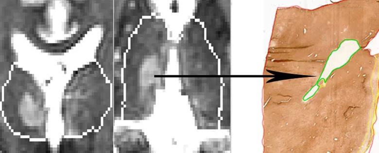
Image content: This image is available to view online.
View image online (https://assets.clevelandclinic.org/transform/fd0fbdbb-42db-456a-9ff7-69602d025820/20-NEU-1882508_Inset_805x325_Mahajan-figure-1_jpg)
Figure 1. The left panels show an MRI of a subject with a sizeable thalamic lesion measuring 1.4 cm3 and a thalamic volume of 18.4 cm3. The expected thalamic volume for the subject’s age is 18.0 cm3, which suggests that even large thalamic lesions do not necessarily correlate with thalamic volume. The right panel shows staining of tissue from the same subject for myelin with proteolipid protein. The demyelinated lesion is outlined in green.
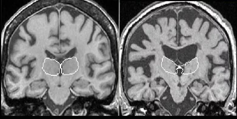
Image content: This image is available to view online.
View image online (https://assets.clevelandclinic.org/transform/5f22114f-5678-4843-9ab7-d8dea97a3d38/20-NEU-1882508_Inset_805x405_Mahajan-fig-2_jpg)
Figure 2. The left MRI is from a subject with one of the greatest thalamic volumes in the study (16.6 cm3), whereas the right MRI is from a subject with one of the lowest thalamic volumes (10.2 cm3) — a 1.6-fold difference. The white matter lesion burden was 2.5-fold higher in the subject on the right versus the one on the left (57.6 vs. 23.4). The white outlines indicate the borders of the thalamus in each subject.
“Our findings suggest that the thalamus is a particular target for neurodegeneration, which is likely because of its extensive connectivity to the rest of the brain,” Dr. Mahajan says. “Axons injured by any number of lesions throughout the brain have a greater likelihood of tracking back to the thalamus compared with other structures.”
One of the most notable aspects of this study was its use of the Cleveland Clinic MS Rapid Brain Donation Program, to which patients with MS have elected to donate tissue after death for use in neuroscience research.
“Although there are a few other MS brain donation programs in the world, ours is unique because of its extensive 24/7/365 collaboration among departments — including our Imaging Institute, Mellen Center and Departments of Pathology, Biomedical Engineering and Neurosciences — and its rapid tissue processing using multiple techniques for high-quality research,” says Dr. Mahajan.
Advertisement
The relationship between lesions and neurodegeneration is of particular interest in MS. Current highly efficacious MS therapies work well in stopping new and enlarging lesions, but they do not help with neurodegeneration directly. The results of the current study suggest that stopping disease activity in early MS by using effective therapies, followed by monitoring with MRI and adjusting therapies if breakthrough activity occurs, will likely impact neurodegeneration by limiting overall lesion burden. Measures such as whole brain and thalamic volumes have seen increasing use in clinical trials in the past few years because of their correlation with neuroperformance measures such as walking speed, manual dexterity, cognition, mood and fatigue.
“Our work supports the involvement of structures such as the thalamus in MS, and we are currently exploring the relationship of individual thalamic subregions with clinical disability,” says Dr. Mahajan. “The hope is that therapeutics may emerge in the not-too-distant future that could repair and decrease the extent of injury within lesions and be ‘neuroprotective,’” he explains, adding that having MRI measures in place to capture neurodegeneration along with lesion volumes will aid in evaluating the efficacy of such therapeutics.
Advertisement
Advertisement

Findings may have implications for understanding the disorders’ pathophysiology

Routine capture of standardized neuroperformance data may expand and refine investigations

Nearly one-fifth of such cases fulfill 2024 McDonald criteria based on biomarkers

Adequate dosing may improve outcomes in well-selected patients, large Cleveland Clinic series suggests

Reimagining the outpatient neurological visit with routine capture of neuroperformance data

Free portal helps researchers classify and share data using the IC-CoDE framework

Large study shows rural patients are less apt to be discharged to inpatient rehab, hampering outcomes

Updates on this fast-evolving therapeutic landscape from a leading trialist