The Case for Including the Central Vein Sign in the Next Diagnostic Criteria for Multiple Sclerosis

Biomarker promises a practical way to reduce misdiagnoses

brain MRI showing central vein sign in a patient with multiple sclerosis

There’s a strong case to be made for incorporating the central vein sign (CVS) into future versions of the McDonald criteria for diagnosis of multiple sclerosis (MS). So contends a recent Viewpoint article in JAMA Neurology by a trio of MS experts from Cleveland Clinic and Cedars-Sinai Medical Center.

Advertisement

Cleveland Clinic is a non-profit academic medical center. Advertising on our site helps support our mission. We do not endorse non-Cleveland Clinic products or services. Policy

“The addition of the CVS to the diagnostic criteria is anticipated to provide increased specificity for the diagnosis of MS and assist clinicians in difficult cases where MRI findings cannot be conclusively determined to be supportive of MS or not,” write Daniel Ontaneda, MD, PhD, and Jeffrey Cohen, MD, both with Cleveland Clinic’s Mellen Center for Multiple Sclerosis Treatment and Research, and Pascal Sati, PhD, of Cedars-Sinai’s Department of Neurology.

These authors argue that better diagnostic tools are needed to reduce the high rate of misdiagnosis of MS, estimated at approximately 20% of cases.

The McDonald criteria specify that MS diagnosis depends on demonstration of dissemination in space (DIS) and dissemination in time (DIT) after a clinical episode characteristic of an MS exacerbation or documented progression. Changes to the criteria over time have allowed for earlier diagnosis, with resulting reductions in specificity. This reduced specificity, together with inappropriate application of MRI criteria and inadequate access to neurological care, is responsible for much of the misdiagnosis of MS, the authors note.

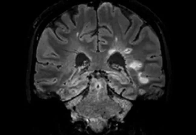
In response, they propose the CVS — i.e., a vein visualized inside a white matter lesion on T2* MRI sequences that appears as a hypointensity relative to the surrounding lesion (see example image above) — as a promising and practice-friendly candidate for helping improve MS diagnosis.

Advertisement

The case for the CVS

The Viewpoint article outlines a number of reasons the CVS shows much promise for real-world use as a diagnostic biomarker:

  • Versatile visualization. The CVS can be visualized at a variety of MRI field strengths and across multiple scanner models.
  • Easy clinical implementation. CVS images can be acquired using commercially available sequences, and acquisition times have been reduced to four to six minutes. Moreover, CVS evaluation does not demand complex image analysis and can be performed by neurologists or neuroradiologists.
  • Effective differentiation from MS mimics. Abundant data support the ability of the CVS to differentiate MS from its common mimics, such as small-vessel disease, migraine and other inflammatory brain disorders.
  • Various rating methods. In addition to the original percentage-based method of rating CVS lesions, abbreviated and straightforward rating methods have been validated and have demonstrated comparable performance, streamlining the rating process. Automated methods of CVS detection have also been developed and will likely be assessed for feasibility.
  • Cost-effectiveness. Additional imaging is not required, as patients already undergo MRI studies with contrast in diagnostic workups.
  • Inclusion of patients with atypical presentations. The CVS can be used as a stand-alone test for patients with atypical presentations of MS — i.e., without a typical clinical event or progression of neurologic disability. “Patients like this currently have no pathway into the McDonald diagnostic criteria,” observes Dr. Cohen, who co-chaired the international panel behind the most recent McDonald criteria revision. “A specific test like the CVS would be clinically valuable in determining the risk of MS.”

Remaining hurdles

Despite these compelling advantages, “validation of the CVS as a diagnostic biomarker still requires some refinement,” the authors write. Several hurdles remain to be cleared:

  • The CVS has not yet been validated in the population where it will likely be most valuable — individuals presenting for an initial diagnosis of MS. To date, most studies have compared patients who have well-established MS with those who have confirmed diagnoses of MS mimics.
  • Although the CVS promises utility for diagnosis of MS with atypical presentations, its performance in this setting requires further study in large, prospective, longitudinal investigations that monitor patients from early in their disease course. The authors note that two such studies are underway. Dr. Ontaneda is co-principal investigator of one of them, CAVS-MS, which includes patients with typical and atypical presentations and is evaluating whether fulfillment of CVS criteria promotes earlier and more accurate diagnosis of MS.
  • The best way of incorporating the CVS into the McDonald criteria is not yet fully clear and will likely be informed by ongoing prospective studies, such as the one profiled here.
  • Most studies of the CVS to date have been done in subspecialty MS centers, so validation of the CVS outside subspecialty centers is needed.

Once these issues are addressed, addition of the CVS to the diagnostic criteria promises to bring the MS community closer to “a simpler and more straightforward diagnosis of MS,” the authors write. They note that its role will likely be refined, however, as data from the necessary prospective studies accumulate.

“We advocate inclusion of the CVS in the McDonald criteria as a necessary step toward a simple, stand-alone MRI characterization of MS without complex diagnostic criteria requirements or the need to demonstrate DIS and DIT,” says Dr. Ontaneda.

Advertisement

The Viewpoint article is available here.

Related Articles

neuron affected by neuromyelitis optica
Novel Monoclonal Antibodies for NMOSD Show Strong Efficacy and Safety in Real-World Study

Early experience with the agents confirms findings from clinical trials

histology image of a gray matter lesion in a multiple sclerosis brain
Study Suggests Protective Role for Microglia at Borders of Gray Matter Lesions in Progressive MS

Findings challenge dogma that microglia are exclusively destructive regardless of location in brain

illustration of a neuron affected by multiple sclerosis
Clinical Trials in Progressive MS: An Assessment of Advances and Remaining Challenges

New review distills insights from studies over the past decade

photo of doctor performing neurologic exam on a patient
A New Guide to the Differential Diagnosis of Multiple Sclerosis

Updated consensus approach helps clinicians efficiently improve diagnostic accuracy

23-NEU-4060108_pregnancy-care-visit_650x450
Reproductive Issues and Multiple Sclerosis: 20 Frequently Asked Questions

The disease is not a barrier to pregnancy, but risks from disease-modifying therapies must be managed

23-NEU-3813861 multiple-sclerosis-patient_650x450
How the SPRINT-MS Follow-On Study Aims to Enhance Future Trials of Progressive Multiple Sclerosis

In search of optimal imaging biomarkers, best methods for long-term follow-up

23-NEU-3991918 CQD hero_650x450
For Older Patients With Multiple Sclerosis, Discontinuing Disease-Modifying Therapy May Be an Option

Risk of new MS activity is small in those older than 55 with stable disease who stop MS therapy

23-NEU-3839389-CQD-Hero-650×450
DELIVER-MS Trial Extended to Guide Philosophy for Long-Term Multiple Sclerosis Therapy

$1.1M award expands follow-up to six years with focus on clinical endpoints

Ad