Locations:

A giant tear of 6 clock hours with a retina that folded on itself
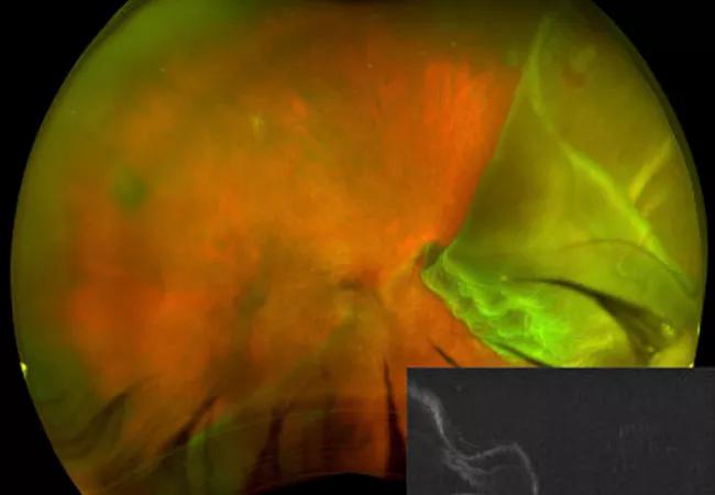
Image content: This image is available to view online.
View image online (https://assets.clevelandclinic.org/transform/67e29b4d-f5f1-43a0-9779-b6665f66deca/17-EYE-1345-Tidal-Wave-CQD-Hero_jpg)
17-EYE-1345-Tidal-Wave-CQD-Hero
By Jeremy A. Lavine MD, PhD; Jaya B. Kumar, MD; Alex Yuan, MD, PhD; and Aleksandra V. Rachitskaya, MD
Advertisement
Cleveland Clinic is a non-profit academic medical center. Advertising on our site helps support our mission. We do not endorse non-Cleveland Clinic products or services. Policy
A 64-year-old man presented at Cleveland Clinic Cole Eye Institute with vision loss in the right eye that had progressed over the course of the previous four weeks. His visual acuity was reduced to hand motion in the right eye and was 20/20 in the left eye. There was no history of trauma.
Wide-field fundus photography (Figure A) showed a giant retinal tear of 6 clock hours. In addition, half of the retina folded on itself, and there was bare choroid superotemporally and a horseshoe tear at 1:30 o’clock. We performed vertical spectral-domain optical coherence tomography, which showed detached macula with the foveal center folded over the inferior retina.
We decided to perform pars plana vitrectomy with silicone oil injection.
At his six-week postoperative visit, the patient’s visual acuity was 20/400 with pinhole. Postoperative fundus photography (Figure B) demonstrated a reattached retina. The vertical optical coherence tomography (OCT) showed silicone oil reflection and ellipsoid zone (arrow) atrophy (between arrowheads) in the foveal center.
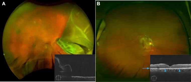
Image content: This image is available to view online.
View image online (https://assets.clevelandclinic.org/transform/a52c953b-c22c-4646-a184-d63e7c2c0d40/17-EYE-1345-Tidal-Wave-CQD-Inset_jpg)
Figure reprinted with permission by Elsevier. Tidal wave retinal detachment, Ophthalmology. 2017 Aug;124(8):1217. doi:10.1016/j.ophtha.2017.01.041.
Rhegmatogenous retinal detachment is a sight-threatening condition. A giant retinal tear is defined as a full-thickness retinal break that extends circumferentially around the retinal periphery for three or more clock hours. Its incidence has been estimated as 1.5 percent of all rhegmatogenous retinal detachments. It affects males more often than females and can occur in both eyes in up to 12 percent of cases.1 In some cases with large giant retinal tears of 6 clock hours or more, the retina can fold on itself.
Advertisement
In the case presented here, the OCT image shows the foveal pit that is not only detached, but also folded over the inferior retina in a “tidal wave” configuration.
Retinal detachments with giant retina tears are usually treated with pars plana vitrectomy and gas or silicone oil as a tamponade agent. Scleral buckle is used in some cases. Re-detachment rates are higher in the presence of giant retinal tears than in cases of retinal detachments due to smaller retinal tears.
Drs. Lavine and Kumar are vitreoretinal fellows at Cole Eye Institute.
Advertisement
Advertisement

The drug does not provide lasting disease control for most patients

New service for patients with diabetes will make annual screening more convenient

Gonioscopy can help determine next steps in reducing IOP

Similarly, patients with noninfectious uveitis have a higher risk of being diagnosed with an autoimmune disease within five years

A review of pathophysiology, risk factors, clinical presentation and treatment options

How computational tools and personalized biomechanics can improve keratoconus detection, ectasia risk assessment and surgical outcomes

Motion-tracking Brillouin microscopy pinpoints corneal weakness in the anterior stroma

Registry data highlight visual gains in patients with legal blindness