Locations:

Prematurity is linked with poorer outcomes of retinal surgery in adulthood
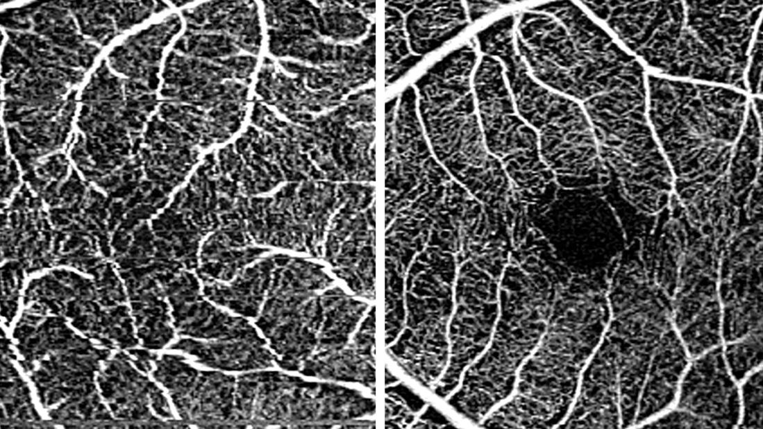
Image content: This image is available to view online.
View image online (https://assets.clevelandclinic.org/transform/7a7455ad-e8a4-4e87-8101-4c5fbb66c7d1/oct-a-pre-term-full-term)
Retina
Adults with a history of preterm birth have a higher risk of glaucoma, retinal tears and detachments, and other eye conditions. A Cleveland Clinic study has found that they also have lifelong changes in macular structure and vascularity — changes that can be detected noninvasively with optical coherence tomography (OCT) and OCT-angiography (OCT-A).
Advertisement
Cleveland Clinic is a non-profit academic medical center. Advertising on our site helps support our mission. We do not endorse non-Cleveland Clinic products or services. Policy
These anatomical differences are apparent even in preterm-birth adults who did not have retinopathy of prematurity (ROP) as an infant.
“Thanks to advances in neonatal care, more babies born prematurely are living full, healthy lives,” says Aleksandra Rachitskaya, MD, a vitreoretinal surgeon at Cleveland Clinic Cole Eye Institute and senior author of the study. “In my own clinic, I have been encountering more adults born preterm. The literature shows that due to anatomical changes in their eyes, they are at higher risk of retinal detachment and needing multiple surgeries for retinal reattachment, compared with adults who were born full term. They also have a higher risk of poorer visual outcome after retinal repair.”
Dr. Rachitskaya recalls one patient in whom she noticed retinal changes during surgery. Those changes made the procedure more challenging. After the surgery, Dr. Rachitskaya learned that the patient had been born prematurely.
“Patients like this were the impetus for our study on preterm-birth adults,” she says. “I wanted to look for biomarkers in this patient population, especially for people not aware of their birth status. Knowing about preterm birth up front could change how we perform surgery on a patient or counsel a patient before surgery.”
The Cole Eye Institute researchers did a retrospective study on 10 premature-birth adults and 11 full-term-birth adults. None of the patients had been treated for ROP as an infant. All had an OCT-A scan at the Cole Eye Institute between 2018 and 2020.
Advertisement
The research team found that premature-birth adults were more likely to have vascular and structural changes of the macula, including:

Image content: This image is available to view online.
View image online (https://assets.clevelandclinic.org/transform/7a7455ad-e8a4-4e87-8101-4c5fbb66c7d1/oct-a-pre-term-full-term)
Left: OCT-A image of an adult with history of premature birth. Pronounced vascularity of the fovea is noted. Right: OCT-A image of an adult with history of full-term birth. Normal avascular fovea is noted.
“Studies of preterm-birth children have found FAZ abnormalities, which seem to correlate with poorer visual acuity,” Dr. Rachitskaya says. “Our study found the same OCT-A changes in preterm-birth adults, including those who had never been treated for ROP and currently have unremarkable fundus examinations. The retinal changes we see in preterm children do continue into adulthood.”
Retinal changes are common in prematurity since blood vessel formation in the peripheral retina typically isn’t complete until around 40 weeks gestation, and foveal development occurs from 25 weeks gestation to after birth.
The Cole Eye Institute study, published in Retina, is among the first, and the largest, to report OCT-A data from preterm adults.
“OCT-A is a nice way to assess vasculature because it does not involve injecting dye, like fluorescein angiography (FA),” Dr. Rachitskaya says. “FA is invasive and time-consuming, and it’s not always comfortable for the patient. We wouldn’t perform FA on a patient unless there is an indication. OCT-A is not invasive and is more comfortable, and it could effectively identify patients born prematurely, who can have abnormal retinal blood vessels.”
Advertisement
For practices that do not have access to OCT-A, CAT and central subfield thickness-to-CAT ratio can be calculated from OCT scans to potentially indicate premature birth in adults.
These OCT and OCT-A findings could be useful in clinical practice, notes Dr. Rachitskaya, who now routinely asks patients about their birth status when assessing them for retinal surgery. She also asks patients who are being evaluated for flashes and floaters, since preterm birth is associated with increased risk of retinal tears.
“Pediatric ophthalmologists are used to asking about this and adjusting the care of patients who were born prematurely,” she says. “Adult practitioners should begin asking about it as well since we will be encountering many more preterm-birth adults as time goes on.”
Patients born prematurely should be educated about the potential differences in their retina and advised on the higher risks of retinal issues.
“We need to establish realistic expectations before surgery and involve the patient in decision-making,” Dr. Rachitskaya says.
Preterm-birth patients may require modified surgical techniques, such as a different positioning of the scleral buckle during repair of retinal detachment.
“Learning how to better manage these patients starts with more effectively identifying who was born preterm,” Dr. Rachitskaya says. “OCT and OCT-A can help us do that, especially in patients who are unaware of their birth status. The next step in our research will be using different imaging modalities to better categorize peripheral changes in these patients.”
Advertisement
Advertisement

Automated diagnosis, risk stratification and treatment assessment are feasible

A look at emerging technology shaping retina surgery

Quality improvement project addresses unplanned extubation

The advanced stage of diabetic retinopathy is among the most challenging for retinal surgeons

How one simple project changed the conversation about care and the patient-parental experience

Target levels of oxygen saturation might only be achieved around one-third of the day, according to available literature

Watch for sudden unilateral vision loss without pain
Levels of volatile organic compounds differ between preterm and full-term infants