March 30, 2017/Pathology

Usual Interstitial Pneumonia: Characteristics and Pathologic Findings

Director of Pulmonary Pathology explains through case study

Identifying usual interstitial pneumonia is more important than ever now that drugs like nintedanib and pirfenidone appear to slow disease progression. Watch Carol Farver, MD, director of Pulmonary Pathology at Cleveland Clinic, explain the characteristics and pathologic findings of this disease through the case of a 65-year-old male:

Advertisement

Cleveland Clinic is a non-profit academic medical center. Advertising on our site helps support our mission. We do not endorse non-Cleveland Clinic products or services. Policy

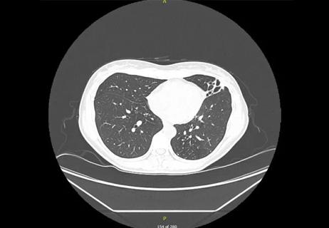
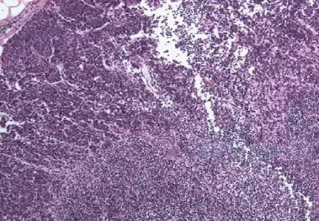
The patient is a white male smoker with a 6 to 8 month history of shortness of breath. The pulmonologist hears crackles and wheezing on exam and orders an imaging study that reveals bilateral basal scarring. A thoracoscopic lung biopsy shows a pathology which, at low power, appears to be a patchy type of fibrosis that does not involve all of the lung.

Advertisement

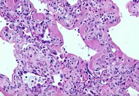
Dr. Farver explains that pulmonary pathologists look for three major characteristics to make the diagnosis of usual interstitial pneumonia. The first is patchy fibrosis which can also be called a temporary heterogenous pattern of injury. The second feature is fibroblasts, which one can see in the interface between the normal and fibrotic lung. When positioned between the scarring and the normal lung, fibroblastic foci are characteristic of usual interstitial pneumonia. The third characteristic is microscopic honeycomb change, which one can see at fairly low power. This patient’s airways had been remodeled to the extent that alveoli were no longer present but rather collagen and fibrosis all around them. Small airways had been remodeled into airways that contained mucus, giving the honeycomb-like appearance characteristic of the severe scarring seen in usual interstitial pneumonia.

This video is part of the Pathology Insights series, featuring important cases, methods and practices personally presented by staff pathologists. The short videos break down information about interesting pathology cases to better inform doctors, laboratory staff, patients or anyone interested in the field of pathology.

Related Articles

21-PUL-2288127 CQD Complex Case 650×450 CT Chest HERO
September 1, 2021/Pathology
Case Study: Refractory Nontuberculous Mycobacterial Infection

Lingulectomy removes infection when antibiotics fail
Tick
May 25, 2021/Pathology/Research
Characterizing Patient Immune Responses to Emerging Tick-borne Virus

Researchers have developed immunoprofiles for an emerging disease with a mortality rate as high as 27%

path
October 23, 2019/Pathology/Case Studies
Vaping Under the Microscope: Pathology of Vaping-Associated Lung Injury

Findings from one of the first published case series

18-PTH-1414-Pleural-Effusion-650×450
December 12, 2018/Pathology/Case Studies
Pleural Fluid: Cyto-Histo Correlation (Video)

A case involving cytology and concurrent biopsy

18-PTH-1299 Mukhopadhyay Hero Image 650x450pxl
August 21, 2018/Pathology/Case Studies
Lung Granulomas Caused by Fungal Infection (Video)

Not all lung nodules are malignant

Ad