Locations:

Staged cavus repair and total ankle arthroplasty
By Sara Lyn Miniaci-Coxhead, MD
Advertisement
Cleveland Clinic is a non-profit academic medical center. Advertising on our site helps support our mission. We do not endorse non-Cleveland Clinic products or services. Policy
For many years, a cavus deformity of more than 10 degrees was considered to be a contraindication for ankle replacement. However, by performing ancillary procedures to correct for the deformities prior to joint replacement, favorable clinical outcomes can be achieved.
When a patient presents with both end-stage osteoarthritis and a cavus deformity, care must be taken to correct malalignment first in order to achieve good outcomes. Failure to balance the ankle joint prosthesis can accelerate wear and premature implant failure. We recently completed six-month follow-up appointments of two patients on whom we performed staged cavus corrections and total ankle replacements—a 61-year-old male and a 62-year-old female. These cases illustrate that though complex, staged cavus corrections with total ankle replacement can be successful with individualized surgical planning.
Cavus foot is a spectrum of foot shapes with abnormally high arches; it can lead to ankle instability, repeated injury and osteoarthritis. It is a fairly common and often subtle deformity, with prevalence estimated at 10% to 25% of the population.
Patient 1 (P1) is a 61-year-old male who presented to our clinic for evaluation of a left cavovarus ankle deformity and arthritis. In the first of two staged operations, we corrected the left cavovarus deformity by performing a dorsiflexion osteotomy of the first metatarsal, peroneal longus to brevis transfer and posterior tendon release, as well as a lateral ligament reconstruction. To prepare the ankle for the arthoplasty, an ankle arthotomy with debridement, and talar cyst was performed. The ankle was pinned in a reduced position. Following this first set of procedures, P1 was placed in a splint. He returned to the clinic two weeks later for splint and suture removal, and was placed in a short leg cast. He was non-weight bearing for six weeks.
Advertisement
Following this initial healing period, we proceeded with the total ankle arthroplasty and syndesmosis fixation. This second stage—the ankle arthroplasty—proceeded without complication. P1 was placed in a well-padded, short-leg splint and was non-weight bearing for three weeks. Sutures were removed at two weeks and P1 was placed in a boot, which was weaned beginning at six weeks post-op.
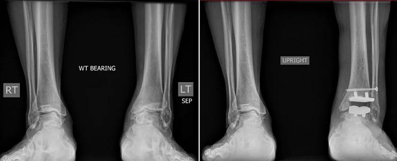
Image content: This image is available to view online.
View image online (https://assets.clevelandclinic.org/transform/84f2b4fa-890c-4a33-8637-5201ef9b0998/19-ORT-1182-Ankle-Replacements-CQD-Inset-1_jpg)
Pre- and post-operative imaging from P1
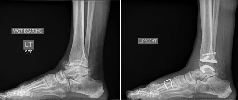
Image content: This image is available to view online.
View image online (https://assets.clevelandclinic.org/transform/2de8a47b-3489-4303-b95b-cf87673fde94/19-ORT-1182-Ankle-Replacements-CQD-Inset-2_jpg)
Pre- and post-operative imaging from P1, lateral view
Patient 2 (P2) is a 62-year-old woman with significant ankle arthritis and recurrent cavovarus deformity. In the first stage of the surgery, to correct the cavus deformity a plantar fascia release, posterior tibial tendon transfer, and calcaneal osteotomy were performed. Again, a lateral ankle ligament reconstruction was also performed. Finally, to prepare the ankle for arthroplasty, the ankle joint was debrided and talar exostectomy was performed. Following this first surgery, P2 was placed in a splint for two weeks, after which time the splint and stitches were removed and her leg placed in a short cast. P2 was non-weight bearing for six weeks.
Six weeks after the first procedure, we removed hardware and osteophytes from the left ankle, followed by total ankle arthroplasty. We noted laxity in the lateral ligaments following osteophyte removal, and again after arthoplasty, which necessitated a repeat lateral ligament repair. P2 was placed in a padded short-leg splint for two weeks, after which time she returned to the clinic for splint and suture removal, and was placed in a tall boot. She began weight bearing as tolerated in the boot at three weeks, but remained in the boot until six weeks post-op, at which time she began a course of physical therapy.
Advertisement

Image content: This image is available to view online.
View image online (https://assets.clevelandclinic.org/transform/ea83d50e-2d3c-4102-83de-f8419412d3af/19-ORT-1182-Ankle-Replacements-CQD-Inset-3_jpg)
Pre- and post-operative imaging from P2
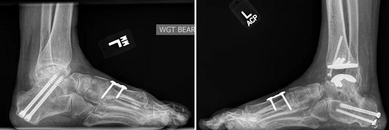
Image content: This image is available to view online.
View image online (https://assets.clevelandclinic.org/transform/9f0d4ad3-ad50-48a9-965e-48f5986b2a42/19-ORT-1182-Ankle-Replacements-CQD-Inset-4_jpg)
Pre- and post-operative imaging from P2, lateral view
Ankle replacement for arthritis in patients with pre-existing ankle deformities often requires multiple procedures to attain a stable, well-balanced prosthesis. At just over six months post-op, both patients are doing well. They both report minimal pain, and have regained ankle range-of-motion.
About the author
Sara Lyn Miniaci-Coxhead, MD, is an orthopaedic surgeon. She specializes in conditions in the foot and ankle.
Advertisement
Advertisement

How it’s similar but different from the direct anterior approach

Collaboration must cross borders and disciplines

Systematic review of MOON cohorts demonstrates a need for sex-specific rehab protocols

Should surgeons forgo posterior and lateral approaches?

How chiropractors can reduce unnecessary imaging, lower costs and ease the burden on primary care clinicians

Why shifting away from delayed repairs in high-risk athletes could prevent long-term instability and improve outcomes

Multidisciplinary care can make arthroplasty a safe option even for patients with low ejection fraction

Percutaneous stabilization can increase mobility without disrupting cancer treatment