Clinical/quantitative model aids in determining surgical candidates

Incorporating clinical automated quantitative MRI measurements into a decision model for temporal lobe epilepsy surgery improves its ability to predict whether freedom from seizures will result. The model is being refined for eventual use in routine clinical practice to guide decisions on the advisability of surgery for individual patients. Details of the model, developed by an international team of epileptologists, were recently published in Brain Communications.
Advertisement
Cleveland Clinic is a non-profit academic medical center. Advertising on our site helps support our mission. We do not endorse non-Cleveland Clinic products or services. Policy
“Despite advances in our ability to evaluate patients for epilepsy surgery, challenges remain in predicting personal outcomes,” says the study’s corresponding author, Lara Jehi, MD, MHCDS, Cleveland Clinic’s Chief Research Information Officer and Director of Outcomes Research in its Charles Shor Epilepsy Center. “This study demonstrates the prognostic value of quantitative MRI beyond the more traditional measures that are taken into account for assessment.”
Predicting outcomes of epilepsy surgery has been a long-term research focus of Dr. Jehi’s. Her team developed a nomogram using basic clinical variables (published in Lancet Neurology), which proved moderately accurate in predicting freedom from seizures, with a C-statistic of 0.6. The C-statistic is an indicator of a model’s quality: a value of 0.5 indicates that it predicts only as well as chance, while 1.0 denotes a perfect model.
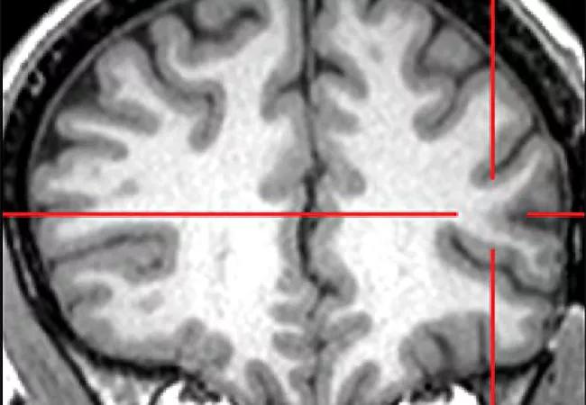
Dr. Jehi’s group conjectured that volumetric brain MRI measures might improve prediction of seizure freedom following epilepsy surgery, as the technology detects abnormalities that may not be visually apparent. Although tools are now available that perform automatic volumetric brain measurements and reference them to matched cohorts, they are not yet readily applicable beyond research settings.
This multicenter retrospective study included 435 patients who underwent temporal lobe surgery for epilepsy between 2010 and 2018 at Cleveland Clinic (n = 289), Mayo Clinic (n = 57) and the University of Campinas, Brazil (n = 89). Median follow-up after surgery was 34 months (interquartile range, 17-60), with a maximum of 116 months.
Advertisement
The team developed multiple prediction models using different data analytic methods and the following clinical factors: preoperative seizure frequency, age at epilepsy onset, age at surgery, duration of epilepsy, sex, etiology, side of surgery, presence of generalized tonic-clonic seizures, MRI abnormalities and type of surgery. Separate models were run with primary endpoints of achieving either freedom from seizures or Engel I at last follow-up.
Presurgical quantitative MRI data were next incorporated into these models to determine whether they improved prediction. The FDA-approved NeuroQuant® software package was used for MRI volumetry.
Findings included the following:
Dr. Jehi notes that the findings were robust, as evidenced by their similarity among the multiple analytic methods the researchers employed.
Dr. Jehi highlights several points learned from these investigations:
Advertisement
Dr. Jehi leads cutting-edge research in prediction models related to epilepsy and recently reviewed the role of algorithms and statistical models in medical decision-making (Epilepsia. 2021 Mar; 62[Suppl 2]:S71-S77). Her team has created models related to long-term outcomes of reoperations (Epilepsia. 2020 Mar;61[3]:465-478), predicting verbal memory decline after temporal lobe resection (Neurology. 2021 May;97[3]:e263-e274), basic scalp EEG findings as a predictor of surgery outcomes (Epilepsia. 2020 Aug 2) and baseline cortical volume loss as a predictor of frontal lobectomy outcomes (Epilepsia. 2021 May;62[5]1074-1084).
“With the increased availability of clinical software packages for automated volume measurements, these measurements can be incorporated into routine clinical practice,” Dr. Jehi concludes. “This current investigation moves us another step towards a meaningful and practical tool to be used by any institution, not just research centers.”
Advertisement
Advertisement

Aim is for use with clinician oversight to make screening safer and more efficient

Rapid innovation is shaping the deep brain stimulation landscape

Study shows short-term behavioral training can yield objective and subjective gains

How we’re efficiently educating patients and care partners about treatment goals, logistics, risks and benefits

An expert’s take on evolving challenges, treatments and responsibilities through early adulthood

Comorbidities and medical complexity underlie far more deaths than SUDEP does

Novel Cleveland Clinic project is fueled by a $1 million NIH grant

Tool helps patients understand when to ask for help