Locations:

Patients frequently have problems with depth perception and interocular suppression
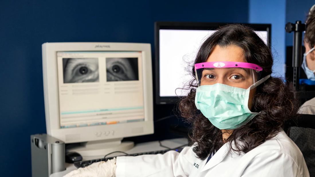
Image content: This image is available to view online.
View image online (https://assets.clevelandclinic.org/transform/794f461b-96d1-4412-bee1-ba5b5ee7cd06/EYE_1994721_10-22-20_050_MLC-jpg)
Dr. Ghasia
Once thought to be a monocular vision condition, amblyopia is increasingly recognized as a binocular disorder that can impact overall vision function. Additionally, patients with amblyopia can have instability of gaze (fixation instability) due to altered fixation eye movements (FEMs) and nystagmus.
Advertisement
Cleveland Clinic is a non-profit academic medical center. Advertising on our site helps support our mission. We do not endorse non-Cleveland Clinic products or services. Policy
Because patient response to amblyopia treatment can vary widely, researchers at Cleveland Clinic Cole Eye Institute analyzed how FEM abnormalities in amblyopia affect binocular visual functions, such as interocular suppression and stereopsis.
“We know that patients with amblyopia have problems with depth perception, but now we also recognize that they frequently experience interocular suppression, which may contribute to the visual acuity deficit of the amblyopic eye,” says Fatema Ghasia, MD, a pediatric ophthalmologist at the Cole Eye Institute.
“There is a spectrum of visual function abnormalities in amblyopia, yet visual functions are not affected to the same extent in each patient. We wanted to evaluate eye movements to see if there’s a pattern of visual function deficit observed in patients with amblyopia,” says Dr. Ghasia, who presented the study at the Association for Research in Vision and Ophthalmology (ARVO) 2022 meeting.
Most of the study’s participants — 34 with amblyopia and seven without — were children. Their eye movements were recorded with infrared video-oculography while viewing with their amblyopic eye, their fellow eye and both eyes.
Researchers analyzed participants’ eye movement traces and categorized amblyopic patients into two groups: those with nystagmus and those without. They further evaluated the eye movement traces to see if participants with nystagmus had fusion maldevelopment nystagmus (FMN), a signature eye movement abnormality that suggests amblyopia will develop in early life, perhaps during infancy.
Advertisement
“Sometimes you can see FMN clinically, but there are plenty of times you need high-resolution eye recordings to pick up subtle nystagmus that may not be obvious,” explains Dr. Ghasia.
Interocular suppression measurement was the primary element of the experiment. Rather than employing the tests used for clinical measurements, researchers used a psychophysical paradigm involving a 3D LCD monitor and polarized glasses. This allowed them to present a different stimulus to each eye to see how the brain perceived it.
“We presented noise dots to the fellow eye and signal dots to the amblyopic eye, and patients were asked to indicate the direction in which the signal dots were moving,” says Dr. Ghasia. “If they could perceive what they were seeing with their amblyopic eye, they would be able to accurately identify the direction of the dots.”
In addition, the team used the Titmus fly test to measure participants’ stereoacuity. They found that patients with FMN who received traditional amblyopia treatment could potentially improve their ability to read an eye chart yet still have poor depth perception. Patients who did not have FMN had stereopsis abnormalities that correlated with their visual acuity abnormalities.
Researchers found that patients with nystagmus and those with FMN were more likely to have abnormal binocular visual functions.
“We learned that studying eye movement recordings in addition to looking at the clinical type of amblyopia helps us better understand the pattern of visual function deficits,” says Dr. Ghasia. “It also may give us a better idea of a patient’s prognosis.”
Advertisement
Regarding FMN, Dr. Ghasia says the tendency is to consider treatment successful when the patient’s vision is 20/20. However, patients who have abnormalities in depth perception or those who are actively suppressing the input from one eye still will have issues with vision quality. Their everyday visual motor tasks can be affected.
“Now that we have identified these patterns, we may need to reconsider our endpoint for improvement in visual acuity of the amblyopic eye,” says Dr. Ghasia. “We may need to consider other aspects of vision, like contrast sensitivity, suppression and stereopsis.”
As eye tracking apps become more widespread, Dr. Ghasia and her team are investigating one app’s utility compared to standard eye trackers. Their hope is that eye tracking will be able to bridge the gap for younger children who are unable to reliably explain what they see.
“Eye movement recordings can play a role because you can have a toddler sit and watch a cartoon character while you capture their eye movements and see if they have an unstable fixation,” says Dr. Ghasia.
This would give pediatricians a better sense of when and when not to refer kids to an ophthalmologist. It also could help guide ophthalmologists on when to start treatment.
“The sooner you detect and treat amblyopia, the better the long-term outcomes,” Dr. Ghasia concludes.
Advertisement
Advertisement

The drug does not provide lasting disease control for most patients

New service for patients with diabetes will make annual screening more convenient

Gonioscopy can help determine next steps in reducing IOP

Similarly, patients with noninfectious uveitis have a higher risk of being diagnosed with an autoimmune disease within five years

A review of pathophysiology, risk factors, clinical presentation and treatment options

How computational tools and personalized biomechanics can improve keratoconus detection, ectasia risk assessment and surgical outcomes

Motion-tracking Brillouin microscopy pinpoints corneal weakness in the anterior stroma

Registry data highlight visual gains in patients with legal blindness