It’s the first step toward reliable screening with your smartphone

Image content: This image is available to view online.
View image online (https://assets.clevelandclinic.org/transform/83301cbd-5afd-42f6-acf5-b3de02580faa/ai-detect-fixational-eye-movement)
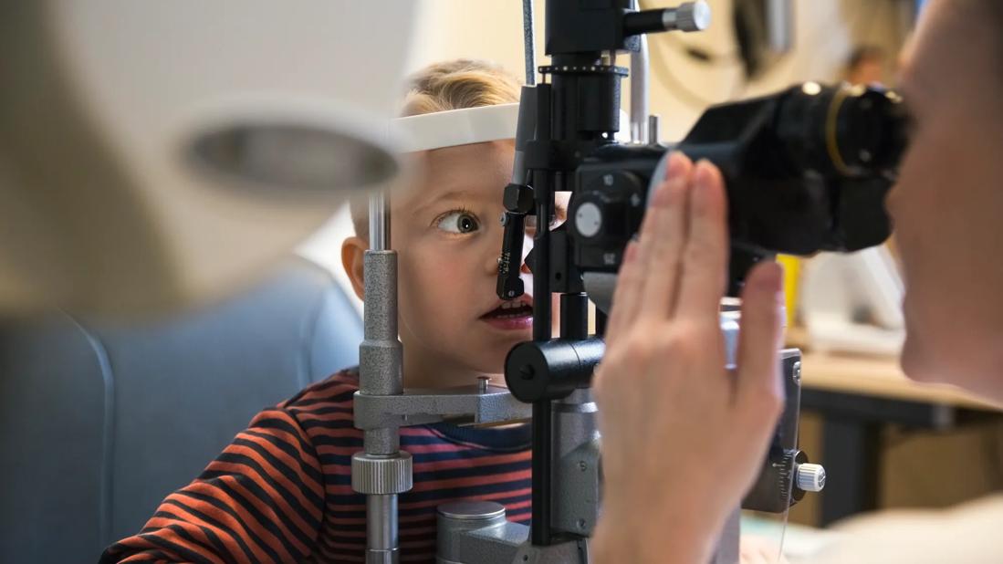
Child getting an eye exam
Fixational eye movement (FEM) may be the key to more accurately and efficiently detecting amblyopia, especially in patients too young to read an eye chart. Now researchers have developed and tested an artificial intelligence (AI) model that effectively distinguishes between the FEM of people with and without lazy eye.
Advertisement
Cleveland Clinic is a non-profit academic medical center. Advertising on our site helps support our mission. We do not endorse non-Cleveland Clinic products or services. Policy
This work builds on a recent study at Cleveland Clinic Cole Eye Institute that found that people with normal vision, regardless of age, have similar FEM patterns in both eyes. This symmetry is not observed in people with amblyopia.
There are three categories of amblyopia:
According to Fatema Ghasia, MD, a pediatric ophthalmologist at the Cole Eye Institute, there are two general ways to screen for amblyopia. The gold standard is visual acuity screening, during which the patient reads an eye chart, one eye at a time, to determine if their vision is different in the right versus the left eye. For young patients who can’t yet read a chart, there is instrument-based screening, such as photoscreening, in which ophthalmologists look for ocular defects in photographs.
“The problem with photoscreening is setting the criteria to get reliable results,” Dr. Ghasia says. “The shape of the eye elongates as people age, so there’s debate about what is ‘normal’ for specific age groups. Companies that make these screening devices set strict standards, but that produces low specificity and a lot of false positives. Many patients referred to us because of abnormal photoscreening actually do not have an eye disorder.”
Detecting abnormality based on eye movement has higher specificity, she says. And portable eye-tracking devices are becoming more widespread. Even newer smartphones and mobile tablets have eye-tracking capabilities.
Advertisement
“Having an AI model could help more accurately screen for amblyopia using these common, affordable tools,” Dr. Ghasia says. “We potentially could detect the disease in patients younger than age 3, long before they can perform visual acuity screening. Because visual system plasticity decreases as people age, there is value in starting treatment for amblyopia as soon as possible. The earlier the treatment, the better the outcomes.”
An observational study recently published in Ophthalmology Science found that building an AI tool to detect amblyopia is feasible.
A research team studied a group of Cole Eye Institute patients, 95 with some type of amblyopia and 40 with normal vision. Each patient sat in a dark room, with their head in a chin rest, and looked at a target on a monitor for 45 seconds. A high-resolution video-based eye tracker recorded how their eyes responded in different viewing conditions (i.e., when using both eyes, the right eye alone or the left eye alone).
The data collected on each patient’s FEM helped train an AI model to distinguish between people with and without amblyopia.
“We gave the raw data to the model and told it whether the data came from an amblyopic eye or healthy eye,” says Dr. Ghasia, senior author of the study. “People with normal vision have similar FEM in both eyes. They have good fixation stability during binocular viewing and slightly less stability during monocular viewing. That instability is much more exaggerated in someone with amblyopia. The AI model caught on to these details.”
Advertisement
Next, the team evaluated the accuracy of the AI model by how well it classified patients according to amblyopia type, amblyopia severity and presence of nystagmus. The model’s accuracy results are presented in the table below.
| Patient subgroup | Accuracy |
|---|---|
| Anisometropia (in which FEM abnormalities are more severe in the amblyopic eye during both binocular and monocular viewing) | 64% |
| Strabismus (in which FEM abnormalities are more severe in the amblyopic eye during binocular viewing but in the nonviewing eye during monocular viewing) | 74% |
| Mixed amblyopia | 72% |
| Treated or mild amblyopia | 73% |
| Moderate or severe amblyopia | 75% |
| With nystagmus | 76% |
| Without nystagmus | 68% |
| Patient subgroup | |
| Anisometropia (in which FEM abnormalities are more severe in the amblyopic eye during both binocular and monocular viewing) | |
| Accuracy | |
| 64% | |
| Strabismus (in which FEM abnormalities are more severe in the amblyopic eye during binocular viewing but in the nonviewing eye during monocular viewing) | |
| Accuracy | |
| 74% | |
| Mixed amblyopia | |
| Accuracy | |
| 72% | |
| Treated or mild amblyopia | |
| Accuracy | |
| 73% | |
| Moderate or severe amblyopia | |
| Accuracy | |
| 75% | |
| With nystagmus | |
| Accuracy | |
| 76% | |
| Without nystagmus | |
| Accuracy | |
| 68% |
“As expected, the model performed slightly better when there was nystagmus, strabismus or more severe disease, but it also was able to detect more inconspicuous amblyopia because of other abnormal eye movements,” Dr. Ghasia says. “Accuracy was quite good in classifying patients regardless of amblyopia type, severity or presence of nystagmus.”
This test AI tool has a promising future, according to Dr. Ghasia. First, however, more study is needed to:
“We’re working on grants to fund these next steps,” Dr. Ghasia says. “I started this work six years ago, when eye tracking wasn’t widely available. Now that eye-tracking technology is becoming more prevalent, I’m excited to see what we can do. If this tool is valuable for diagnosing amblyopia, it also may be valuable for detecting neurodegenerative and mental health conditions associated with asymmetrical interocular movements.”
Advertisement
This research was supported by multiple grants, including the Research to Prevent Blindness Disney Amblyopia Award, Research to Prevent Blindness International Research Collaborators Award and the Lerner Research Institute Artificial Intelligence in Medicine grant.
Advertisement
Advertisement
Registry data highlight visual gains in patients with legal blindness
Study is first to show reduction in autoimmune disease with the common diabetes and obesity drugs
Fixational eye movement is similar in left and right eyes of people with normal vision
Oral medication may have potential to preserve vision and shrink tumors prior to surgery or radiation
A new online calculator can determine probability of melanoma
Only 33% of patients have long-term improvement after treatment
Novel collaborative approach helps patient avoid orbital exenteration
Interventions abound for active and stable phases of TED